Hydrogel, Gel/Plastic and Rubbery Clots

Hydrogel, Gel/Plastic and Rubbery Clots
Are they all the same ‘thing’?

by David Nixon, Nixonlab
March 2, 2024


Hi Y’all,

I think that the microscopic findings of hydrogel and structures, the macroscopic finding from a sample of live blood that shows an abnormal protein ‘gel/plastic’ and the rubbery white clots that have been found by embalmers for over two years are all showing the same pathology.

They all showed up at the same time, although the gel/plastic was not recognised until later I bet it would have been there if anybody had looked earlier.

In all three situations I believe we are dealing with proteins that were soluble within hydrogels but have become insoluble and ‘drop’ out of solution.

We are all familiar with adding hot water to a sheet of gelatin and forming jelly.


Simplistically we are adding heat and forming a hydrogel. Imagine if we extracted heat and produced bits of gelatin sheet.

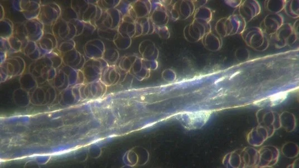
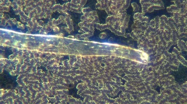
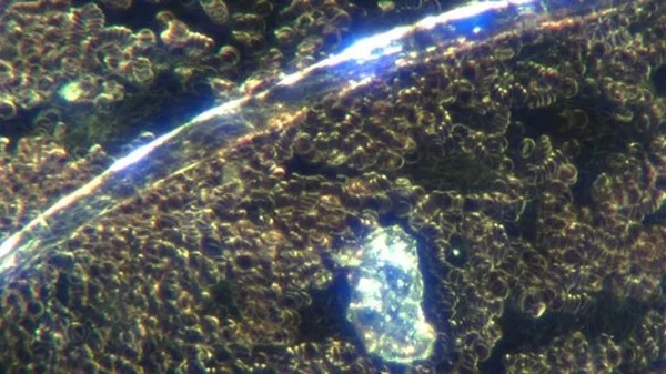
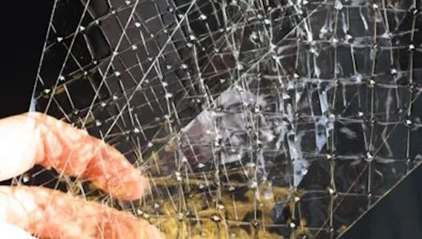
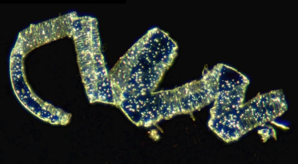
I think this, or something similar, is what we are observing microscopically. I don’t think the structure below is in the blood in the finger but it develops from the time of the finger prick to the time of viewing. This is a darkfield image, photo-stitch, with 40x objective. I have seen structures with this texture on a few occasions but not often. Clearly there are large amounts of nanoparticles present. I had to turn the gain down which has meant the surrounding red blood cells have largely disappeared.


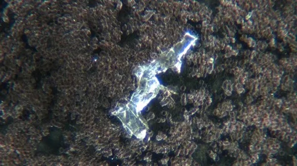
Macroscopically, the identified abnormal protein that was identified and investigated by Prof Arne Burkhart we have call ‘gel/plastic’, clearly it is not a gel or plastic but this name serves as a place sitter. Ron Norris and Michael Merrick and more recently Bill from Missouri have led the way with drawing their own blood and centrifuging it as per Ron (please see Ron’s Substack). I have trialled successfully (and this is ongoing) Mike Merrick’s protocol for the oral use of thieves’ oil (LAC microscopy on Telegram). Unfortunately have not made time to write this up but will do so asap, promise!

Lastly, thoughts on the rubbery clots. I think these are mainly protein and form from the hydrogels after death, at least in the main part and I don’t think that they are what is causing death per se. But clearly they are associated with what’s going on.

So what do we do? Minimise EMF exposure. Avoid processed food. Ensure that drinking water is either distilled (then restructured and re-mineralised) or been through a reverse osmosis filter or is from a trusted non-contaminated source. Take regular activated charcoal, sodium citrate and follow the discussions with regard to gel/plastic. Consider other supplements from trusted sources. Consider flu-like symptoms as signs of toxicity. Don’t stick anything up your nose. Masks don’t help.


Connect with David Nixon


What’s in the Blood?

What’s in the Blood?


“We can gnash our collective teeth in frustration and angst at our political leaders and public health officials but surely the presence of these abnormalities in the blood indicates a titanic problem and hints, strongly I believe, towards a more sinister agenda. We must look and we must act on what we see.”

~ Dr. David Nixon


by David Nixon, Nixonlab
originally published February 24, 2023


What’s in the blood?

“For every mistake made for not doing: ten are made for not looking”

(Poster on wall at Otago Medical School circa 1987 – Author unknown)

Live blood analysis has a history longer than vaccination. It is interesting to note that proponents of both were on opposite sides of the “debate”. It was the work of French scientists Antoine Bechamp and Claude Bernard and later that of Professor Günther Enderlein that advanced the use of the microscope and challenged the medical establishment of the day. That challenge has never gone away.

Looking at blood under a microscope would seem like a fairly sensible thing to do given the significant concerns that exist amongst experienced, usually non-medically trained practitioners. Unfortunately, promoting live blood analysis as a useful and complementary modality is akin to questioning vaccine science. Don’t do it. Don’t do either when it comes to that.

In Australia in the early 2000’s Metagenics ran training programmes for general practitioners, naturopaths and other health practitioners. Unfortunately, they stopped providing this training due to increasing compliance costs and the number of practitioners providing live blood analysis has reduced significantly.

However, in late 2022 when questioning the safety and effectiveness of a vaccine that is “safe and effective” it would seem sensible to at least have another look, especially given the increasing rhetoric “there is nothing to see here.” This is of course a medical-centric comment because live blood analysis has been offered continuously for decades and many practitioners are highly experienced in providing and interpreting this investigation.

These practitioners are exceptionally alarmed about the changes and the abnormal structures that have been seen in the blood that are now visible in both vaccinated and unvaccinated patients. These structures were not seen before late 2021 – in anybody. These practitioners are increasingly marginalised from the health care machine. They do not have an effective voice. A bit like the patients that are suffering from clear vaccine related injury. It will be a historical outrage that the vaccine injured patients have been treated so poorly.

Unpublished data from a researcher in Australia has shown rouleaux and or thrombi being present >6 months after the last vaccine dose in >81% of vaccinated samples vs 16% of unvaccinated samples in a blinded analysis (n=314). One reason that this data is unpublished is because of the effort and cost required to gain ethics approval for conducting this study. Ethics approval that the current Covid-19 vaccination programme has not had to obtain.

In a recently published Italian study Cipelli and colleagues noted that more than 94% of 1006 symptomatic patients within 1 month of vaccination with Pfizer or Moderna had significant abnormalities in the blood. Furthermore 12 patients were checked before and after vaccination and showed normal appearances prior to vaccination and similar changes to the the other patients after vaccination.

Similar findings to the patients in this study have been observed in blood of vaccinated patients all around the world. Typically, these take the form of red blood cell stacking, referred to as rouleaux, evidence of metabolic stress and hypercoagulability and abnormal inclusions which includes ribbons, tubular structures and crystals.

A thicker tubular structure at 400x magnification with 2 neutrophils attached.

A similar structure with marked rouleaux in the background.

A large folded ribbon structure, rouleaux and fibrin deposition.

A tubular structure and crystal with blood again showing rouleaux and fibrin deposition.

We can debate the origin of Covid-19 and the subsequent global response.  We can gnash our collective teeth in frustration and angst at our political leaders and public health officials but surely the presence of these abnormalities in the blood indicates a titanic problem and hints, strongly I believe, towards a more sinister agenda. We must look and we must act on what we see.

David Nixon Nov 2022


Connect with Dr. David Nixon


Pfizer Covid Vax Under the Microscope: Dark Field Microscopy by Dr. David Nixon

Pfizer Covid Vax Under the Microscope: Dark Field Microscopy by Dr. David Nixon


Dark Field Microscopy by Dr David Nixon

by Dr. David Nixon
source: Team Enigma

November 3, 2022

What we know is explained in the video.


Pfizer Vials Under Microscope – Structures Grow in Electromagnetic Field.

by Sasha Latypova, Due Diligence podcast

October 17, 2022


[Video source: Team Enigma]

I recently spoke with Dr. David Nixon who was able to conduct several experiments with Pfizer injectable substance under standard optical microscope. David found that when the sample is shielded from electromagnetic field by the Faraday bag no assembly happens in it. When the slide is left overnight near a wifi router, square shaped structures appear, and then continue to grow for a long period of time.


Connect with Dr. David Nixon

Connect with Sasha Latypova — substackBitChute channel


See related:

What’s Inside the Jabs? The Truth Continues to Emerge