It seems surprising, even ironic, to conclude a health food cookbook, in which we have warned against sugar, yeasted foods and tea, with a tonic made from sugar, yeast and tea! But the kombucha “mushroom” (which is actually a symbiotic colony of yeast and bacteria) acts on sugar and tea to produce not only acetic and lactic acid but also small amounts of a potent detoxifying substance, glucuronic acid. Normally this organic acid is produced by the liver in sufficient quantities to neutralize toxins in the body— whether these are naturally produced toxins or poisons ingested in food and water. However, when liver function becomes overloaded, and when the body must deal with a superabundance of toxins from the environment—certainly the case with most of us today—additional glucuronic acid taken in the form of kombucha is said to be a powerful aid to the body’s natural cleansing process, a boost to the immune system and a proven prophylactic against cancer and other degenerative diseases.
More importantly, kombucha is the cure for a hot day—it tastes delicious and refreshing. A fizzy, dark colored, energizing beverage, at the same time acidic and slightly sweet, this gift to the world from the Ural mountain region of Russia qualifies as the soft drink of the twenty-first century, the answer to the scourge of cola drinks that now wreaks havoc with the health of Western populations.
Remove from heat, add the tea bags and allow the tea to steep until water has completely cooled.
Remove tea bags. Pour cooled liquid into a 4-quart pyrex bowl and add ½ cup kombucha from previous batch.
Place the mushroom on top of the liquid.
Make a crisscross over the bowl with masking tape, cover loosely with a cloth or towel and transfer to a warm, dark place, away from contaminants and insects. [Alternatively, put a loose cloth over the container and tie it in place so it dosen’t fall in!]
In about 7 to 10 days the kombucha will be ready, depending on the temperature.
It should be rather sour and possibly fizzy, with no taste of tea remaining.
Transfer to covered glass containers and store in the refrigerator. (Note: Do not wash kombucha bowls in the dishwasher.)
When the kombucha is ready, your mushroom will have grown a second spongy pancake. This can be used to make other batches or given away to friends.
Store fresh mushrooms in the refrigerator in a glass container—never plastic. A kombucha mushroom can be used dozens of times. If it begins to turn black, or if the resulting kombucha doesn’t sour properly, it’s a sign that the culture has become contaminated. When this happens, it’s best to throw away all your mushrooms and order a new clean one.
Note: White sugar, rather than honey or Rapadura, and black tea, rather than flavored teas, give the highest amounts of glucuronic acid. Non-organic tea is high in fluoride so always use organic tea.
A word of caution: Some individuals may have an allergic reaction to kombucha. If you have allergies, start with a small taste to observe any adverse effects. If you react badly, use beet kvass several weeks to detoxify and then try again.
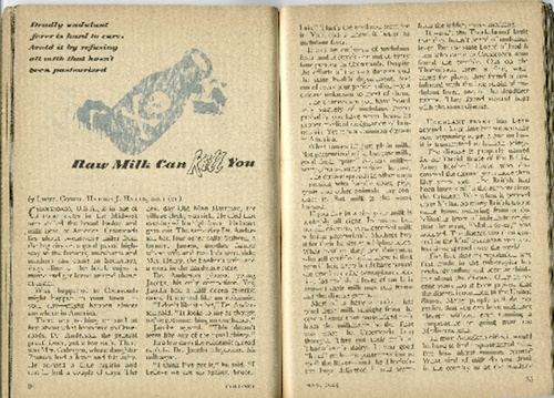
Few of us were born when the forces for milk pasteurization launched the first major attack on Nature’s perfect food. In 1945, a magazine called Coronet published an article, “Raw Milk Can Kill You,” blaming raw milk for an outbreak of brucellosis in a town called Crossroads, U.S.A., killing one-third of the inhabitants. The Reader’s Digest picked up the story and ran it a year later.
Just one problem with this piece of “reporting.” There was no town called Crossroads and no outbreak of brucellosis. The whole story was a fabrication—otherwise known as a lie. And lies about raw milk have continued ever since.
Unfortunately, the fictitious Crossroads story paved the way for laws against selling raw milk, starting with Michigan in 1948.
Here’s another example of lies against raw milk (which I referenced in an earlier post,[i] but it is worth repeating). In 2007, John F. Sheehan, BSc (Dy), JD, US Food & Drug Administration, Center for Food Safety & Applied Nutrition (USFDA/CFSAN), Division of Dairy and Egg Safety, prepared a Powerpoint maligning raw milk; it was presented to the 2005 National Conference on Interstate Milk Shipments (NCIMS) by Cindy Leonard, MS.[ii]
As shown in the table below, all of the fifteen reports associating outbreaks of foodborne illness with raw milk that Sheehan cites are seriously flawed. For example, in two of the fifteen, the study authors presented no evidence that anyone consumed raw milk products and in one of them, the outbreak did not even exist. Not one of the studies showed that pasteurization would have prevented the outbreak.
No Valid Positive Milk Sample
12/15
80%
No Valid Statistical Association with Raw Milk
10/15
67%
Findings Misrepresented by FDA
7/15
47%
Alternatives Discovered, Not Pursued
5/15
33%
No Evidence Anyone Consumed Raw Milk Products
2/15
13%
Outbreak Did Not Even Exist
1/15
13%
Did Not Show that Pasteurization Would Have Prevented Outbreak
15/15
100%
Fast forward to the present and the ruckus about bird flu in dairy cows—more lies, very clever lies, but lies nevertheless.
In a press release dated March 25, 2024 ,[iii] the U.S. Department of Agriculture (USDA), Food and Drug Administration (FDA) and Centers for Disease Control and Prevention (CDC), as well as state veterinary and public health officials, announced investigation of “an illness among primarily older dairy cows in Texas, Kansas, and New Mexico that is causing decreased lactation, low appetite, and other symptoms.”
The agencies claim that samples of unpasteurized milk from sick cattle in Kansas and Texas have tested positive for “highly pathogenic avian influenza (HPAI).” Officials blame the outbreak on contact with “wild migratory birds” and possibly from transmission between cattle. The press release specifically warns against consumption of raw milk, a warning repeated in numerous publications and Internet postings.
According to the press release, national laboratories have confirmed the presence of HPAI (Highly Pathogenic Avian Influenza) through testing, but it does not reveal the type of test used to detect this so-called viral illness.
THE FIRST LIE: Researchers have found HPAI virus in the milk of sick cows.
Officials have NOT found any viruses in the milk or any other secretions of the sick cows. The CDC has yet to reply to repeated requests for proof of finding the isolated HPAI virus in any fluid of any sick chicken or other animal.[iv] Nor have health and agriculture agencies in Canada,[v] Japan[vi], the UK[vii] and Europe[viii] provided any proof of an isolated avian influenza virus.
As for all the studies you can find in a PubMed search claiming “isolation” of a virus, not one of them shows the true isolation of a virus, any virus, from the fluids (phlegm, blood, urine, lung fluids, etc) of any animal, bird or human.[ix]
The truth is that “viruses” serve as the whipping boy for environmental toxins, and in the confinement animal system, there are lots of them–hydrogen sulfide, carbon dioxide, methane and ammonia from excrement, for example.[x] Then there are toxins in the feed, such as arsenic added to chicken feed, and mycotoxins, tropane and β-carboline alkaloids in soybean meal.[xi] By blaming nonexistent viruses, agriculture officials can avoid stepping on any big industry toes nor add to the increasing public disgust with the confinement animal system.
Way back in 2006, researchers Crowe and Englebrecht published an article entitled, “Avian flu virus H5N1: No proof for existence, pathogenicity, or pandemic potential; non-‘H5N1’z causation omitted.”[xii]Nothing has changed since then.
Here’s your homework assignment: Contact USDA at Aphispress@usda.gov and ask them to provide proof of the isolation of the HPAI virus or any virus in the milk of the sick cattle.
SECOND LIE: National laboratories have confirmed the presence of HPAI (Highly Pathogenic Avian Influenza) through testing.
They don’t say anything about the kind of test they used, but it almost certainly the PCR (polymerase chain reaction) test. The PCR test detects genetic material from a pathogen or abnormal cell sample and allows researchers to make many copies of a small section of DNA or RNA. The test was not designed to determine or diagnose disease, it was designed to amplify or increase a certain piece of genetic material.
Each “amplification” is a doubling of the material. If you amplify thirty times you will get a negative; amplify 36 times or more, and you will get a positive. At 60 amplifications, everyone will “test positive” for whatever bit of genetic material you believe can cause disease.[xiii] If you want to show that you have a pandemic brewing, just amplify, amplify, amplify. Folks, this is not a valid test, not good science by any stretch of the imagination—especially as there was no virus to begin with.
How many times did our health officials amplify the samples they obtained from the milk of the sick cows? Be sure to ask them when you email Aphispress@usda.gov for proof of the virus.
THIRD LIE: The “virus” is highly pathogenic.
According to the Wall Street Journal, one—just one–person working in the dairies got sick and tested positive for avian influenza after exposure to dairy cattle presumed to be infected with the H5N1 bird flu.[xiv] The person reported eye redness, or conjunctivitis, as his only symptom—a symptom that can be explained by exposure to any of the many airborne toxins in confinement dairies. (How are they treating the illness? With vitamin A and herbal eyedrops? No, the poor sod is getting treatment with a toxic antiviral drug.)
According to the CDC, the disease in humans ranges from mild infections, which include upper-respiratory and eye-related symptoms, to severe pneumonia. If the “virus” is so highly pathogenic, we’d expect a lot of workers working around these sick cows to end up in the hospital. . . but we’ve heard of none so far.
FOURTH LIE: You can get avian fly from drinking raw milk, but pasteurized milk is safe
According to medical biologist Peg Coleman,[xv] “Recent risk communications from CDC, FDA, and USDA regarding transmission of highly pathogenic avian influenza virus or HPAI (subtype H5N1) to humans via raw milk include no supporting evidenceof viral transmission from raw milk to humans in the peer-reviewed literature. . . An extensive body of scientific evidence from the peer-reviewed literature . . . does not support the assumption by these US government agencies that [non-existent] HPAI transmits to humans via milkborne or foodborne routes and causes disease. Nor does the scientific evidence support the recommendation that consumers should avoid raw milk and raw milk products [emphasis in the original].”[xvi]
Coleman notes the suite of bioactive components in raw milk, including bovine milk, that destroy pathogens and strengthen the gut wall. “Many of these bioactive components of raw milk are . . . sensitive to heat and may be absent, inactive, or present in lower concentrations in pasteurized milks. . . Cross-disciplinary evidence demonstrates that raw milk from healthy cows is not inherently dangerous, consistent with the CDC evidence of trends for 2005-2020 and evidence of benefits and risks. There is no scientific evidence that HPAI in raw milk causes human disease.”
And while USDA, FDA and CDC assure the public that pasteurization will make milk safe, they note that “Milk from infected animals is being diverted or destroyed,” implying that pasteurization alone does not guarantee safety. In any event, sales of industrial pasteurized milk continue their relentless decline.
Fortunately, raw milk drinkers are already skeptical of government pronouncements and are skilled at seeing through lies. Both large and small raw milk dairy farms report that sales are booming. The current bird flu fracas is just another Crossroads, U.S.A., a bunch of lies fostered by a dishonest dairy industry taking aim at the competition.
by Sally Fallon Morell, Nourishing Traditions, Weston A. Price Foundation
January 11, 2022
With the Covid vaccination program now in shambles, officials are focusing their fear porn on the measles, as evidenced by a December 27, 2022 front page article appearing in the Washington Post.
“Diseases resurging as parents resist shots: Outbreaks of measles, chickenpox tied to rise in anti-vaccine sentiment,” places the blame on “parent resistance of routine childhood immunizations. . . intensifying a resurgence of vaccine-preventable diseases.” The article does not provide any information on exactly how many of the children who contracted measles were not vaccinated and how many were, but consists mostly of quotes from hand-wringing public officials about children not getting their shots.
A CBS news report tells a rather different story. In an Ohio outbreak involving eighty-two children, 94 percent of whom were under age five, “all of the children impacted by the outbreak are at least partially unvaccinated, meaning they have only received one dose of the necessary two for the measles-mumps-rubella vaccine, known as MMR, although four children still have an unknown vaccination status. Children are recommended to get their first dose between 12 and 15 months of age and the second between the age of 4 and 6.”
Since most of the Ohio children afflicted were under five, this means that all of them were in fact “fully vaccinated” since the second dose is recommended for children ages four to six. Vaccinated children who get the measles provide proof that measles is not “vaccine-preventable” at all. In fact, we are justified in asking whether children getting the measles so young—normally the illness occurs in children around age seven or eight—is an indication that the vaccine may be causing children to contract the measles too early in life.
The cause of measles, according to public health agencies, is a “highly contagious virus” spread through the coughings and sneezings of the afflicted—or even viruses remaining on surfaces that measles sufferers have touched. The problem is that scientists have been unable to find said virus in these fluids. Credit for the “isolation” or “discovery” of the “agent of measles” goes to John F. Enders, winner of the 1954 Nobel Prize in physiology and medicine.
Enders developed techniques of “propagating” the virus in a culture. The procedure involved taking throat cultures from children sick with measles, mixing them with “sterile fat-free milk,” adding a high dose of penicillin or streptomycin and then centrifuging this goop. The resulting supernatant fluid or sediments were again mixed with milk and used as inocula in different experiments, where they were added to various types of tissue including human kidney, human embryonic lung, human embryonic intestine, human uterus, rhesus monkey testes, human embryonic skin and muscle, human foreskin (!), rhesus monkey kidney and embryonic chick tissue. You can’t accuse Dr. Enders of not being thorough! Only the rhesus kidney cells gave Enders the results he wanted—a breakdown of the cells in the tissues. And yet, the consensus is that animals don’t get measles!
The culture medium consisted of bovine amniotic fluid, beef embryo extract, horse serum, eye of newt and toe of frog. Just kidding about the last two items. To this mixture of biological materials (and they are calling this an “isolation”!) was added phenol red, antibiotics and—strangely—soy trypsin inhibitor. The monkey kidney cells broke down—cell boundaries were obliterated, the nuclei deteriorated and large and small vacuoles (empty spaces) formed. What caused this breakdown? Enders claimed it was the “agent of measles” but a more likely candidate was the antibiotics, especially streptomycin, which is a kidney toxin.
Since Ender’s day, thousands of papers on virus “isolation” have cluttered up the scientific literature, using variations of his technique to claim the pathogenic effects of “viruses,” but Enders paper was unique: it included a control. Enders looked at monkey kidney cells that had not received an inoculation of measles material, and the cells broke down anyway. “The cytopathic changes it induced in the unstained preparations could not be distinguished with confidence from the viruses isolated from measles.” After staining, the measles-cultured cells did look different, with more deterioration of the nuclei, but remember, there were antibiotics in the cultures as well.
Symptoms of measles include a diffuse red rash, high fever, cough, runny nose and red, watery eyes (conjunctivitis), and occasionally abdominal pain, vomiting and diarrhea. These symptoms usually subside in a few days but in malnourished children, measles can result in serious side effects such as blindness or seizures, and even can be fatal.
According to the “experts,” no treatment is available for the measles. Acetaminophen and NSAIDS for pain and fever is a common recommendation, along with bed rest and plenty of liquids.
Actually, there is a treatment for measles, a treatment that can be lifesaving in cases of severe measles: vitamin A. I find it shocking that public health officials can turn their backs on the accumulated science about vitamin A and measles. A literature search turns up over five hundred studies on this subject, such as this one entitled, “Low serum retinol is associated with increased severity of measles in New York City children,” in which the authors concluded “Children with no known prior vitamin A deficiency exhibited a significant decline in their serum retinol levels during the acute phase of measles. This decline in circulating retinol was associated with increased duration of fever, higher hospitalization rates, and decreased antibody titers.”
Or this one: “Vitamin A for the treatment of children with measles–a systematic review,” which begins with the statement, “Vitamin A deficiency is a recognized risk factor for severe measles,” and concludes “that 200,000 IU of vitamin A repeated on 2 days should be used for the treatment of measles as recommended by WHO in children admitted to hospitals in areas where the case fatality is high.”
Recommended by WHO! But you aren’t reading about the miraculous results of vitamin A treatment for measles patients in publications like the Washington Post. Instead, there is the constant push for vaccinations, even though the MMR (measles, mumps, rubella) vaccine can have serious side effects, including autism. Ingredients in the MMR vaccine include chick embryo cell culture, WI-38 human diploid lung fibroblasts, MCR-5 cells, vitamins, amino acids, fetal bovine serum, sucrose, glutamate, recombinant human albumin, neomycin, sorbitol, hydrolyzed gelatin, monosodium L-glutamate, sodium bicarbonate, potassium chloride, potassium phosphate, sodium phosphate and sodium chloride. (Potassium chloride is used to cause cardiac arrest as the third drug in the “three drug cocktail” for executions by lethal injection.)
Instead of vaccinations: let your child get the measles! The treatment is simple: bed rest in a darkened room (to avoid overstimulating the eyes); cold compresses for fever; and cod liver oil (use an eye dropper), smoothies of egg yolk, cream and maple syrup, and a little liver pate for vitamin A. With vitamin A-rich cod liver oil and food, your child will be right in no time, and will have protection against the serious effects of high fever. One school of thought holds that having the measles strengthens the immune system and provides protection against cancer later in life.
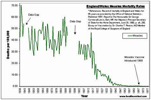
So if it’s not a virus, what causes the measles? Since measles is obviously an effort by the body to detoxify, environmental toxins, especially in the water, are a likely candidate. The decline in measles in industrialized countries, especially deaths from measles, parallels the cleaning up of our cities and cleaner water for everyone. Diets also improved, especially up to the Second World War, when people still drank whole milk, ate butter and took cod liver oil.
Even so, children still get the measles and one theory holds that children go through a natural, even programmed, cleansing as they make the transition from early to middle childhood around age seven. Children with measles may even “communicate” to other children of the same age that it’s time to go through this important process . Certainly not everyone in a household gets the measles when one child has it, not even other children.
Once we throw off the “virus” theory of measles, we can explore the true causes of this and other childhood diseases. Meanwhile, a nutrient-dense diet is the best protection for your child.
Sally Fallon Morell is best known as the author of Nourishing Traditions®: The Cookbook that Challenges Politically Correct Nutrition and the Diet Dictocrats. This well-researched, thought-provoking guide to traditional foods contains a startling message: animal fats and cholesterol are not villains but vital factors in the diet, necessary for normal growth, proper function of the brain and nervous system, protection from disease and optimum energy levels. View all posts by Sally Fallon Morell
Isolation: The action of isolating; the fact or condition of being isolated or standing alone;
separation from other things or persons; solitariness.
– Oxford English Dictionary
The controversy over whether the SARS-CoV-2 virus has ever been isolated or purified continues. However, using the above definition, common sense, the laws of logic and the dictates of science, any unbiased person must come to the conclusion that the SARS-CoV-2 virus has never been isolated or purified. As a result, no confirmation of the virus’ existence can be found. The logical, common sense, and scientific consequences of this fact are:
the structure and composition of something not shown to exist can’t be known, including the presence, structure, and function of any hypothetical spike or other proteins;
the genetic sequence of something that has never been found can’t be known;
“variants” of something that hasn’t been shown to exist can’t be known;
it’s impossible to demonstrate that SARS-CoV-2 causes a disease called Covid-19.
In as concise terms as possible, here’s the proper way to isolate, characterize and demonstrate a new virus. First, one takes samples (blood, sputum, secretions) from many people (e.g. 500) with symptoms which are unique and specific enough to characterize an illness. Without mixing these samples with ANY tissue or products that also contain genetic material, the virologist macerates, filters and ultracentrifuges i.e. purifies the specimen. This common virology technique, done for decades to isolate bacteriophages1 and so-called giant viruses in every virology lab, then allows the virologist to demonstrate with electron microscopy thousands of identically sized and shaped particles. These particles are the isolated and purified virus.
These identical particles are then checked for uniformity by physical and/or microscopic techniques. Once the purity is determined, the particles may be further characterized. This would include examining the structure, morphology, and chemical composition of the particles. Next, their genetic makeup is characterized by extracting the genetic material directly from the purified particles and using genetic-sequencing techniques, such as Sanger sequencing, that have also been around for decades. Then one does an analysis to confirm that these uniform particles are exogenous (outside) in origin as a virus is conceptualized to be, and not the normal breakdown products of dead and dying tissues.2 (As of May 2020, we know that virologists have no way to determine whether the particles they’re seeing are viruses or just normal break-down products of dead and dying tissues.)3
2 “Extracellular Vesicles Derived From Apoptotic Cells: An Essential Link Between Death and Regeneration,” Maojiao Li1 et al, Frontiers in Cell and Developmental Biology, 2020 October 2. https://www.frontiersin.org/articles/10.3389/fcell.2020.573511/full — accessed 2/15/21
3“The Role of Extraellular Vesicles as Allies of HIV, HCV and SARS Viruses,” Flavia Giannessi, et al, Viruses, 2020 May
If we have come this far then we have fully isolated, characterized, and genetically sequenced an exogenous virus particle. However, we still have to show it is causally related to a disease. This is carried out by exposing a group of healthy subjects (animals are usually used) to this isolated, purified virus in the manner in which the disease is thought to be transmitted. If the animals get sick with the same disease, as confirmed by clinical and autopsy findings, one has now shown that the virus actually causes a disease. This demonstrates infectivity and transmission of an infectious agent.
None of these steps has even been attempted with the SARS-CoV-2 virus, nor have all these steps been successfully performed for any so-called pathogenic virus. Our research indicates that a single study showing these steps does not exist in the medical literature.
Instead, since 1954, virologists have taken unpurified samples from a relatively few people, often less than ten, with a similar disease. They then minimally process this sample and inoculate this unpurified sample onto tissue culture containing usually four to six other types of material — all of which contain identical genetic material as to what is called a “virus.” The tissue culture is starved and poisoned and naturally disintegrates into many types of particles, some of which contain genetic material. Against all common sense, logic, use of the English language and scientific integrity, this process is called “virus isolation.” This brew containing fragments of genetic material from many sources is then subjected to genetic analysis, which then creates in a computer-simulation process the alleged sequence of the alleged virus, a so called in silico genome. At no time is an actual virus confirmed by electron microscopy. At no time is a genome extracted and sequenced from an actual virus. This is scientific fraud.
The observation that the unpurified specimen — inoculated onto tissue culture along with toxic antibiotics, bovine fetal tissue, amniotic fluid and other tissues — destroys the kidney tissue onto which it is inoculated is given as evidence of the virus’ existence and pathogenicity. This is scientific fraud.
From now on, when anyone gives you a paper that suggests the SARS-CoV-2 virus has been isolated, please check the methods sections. If the researchers used Vero cells or any other culture method, you know that their process was not isolation. You will hear the following excuses for why actual isolation isn’t done:
There were not enough virus particles found in samples from patients to analyze.
Viruses are intracellular parasites; they can’t be found outside the cell in this manner.
If No. 1 is correct, and we can’t find the virus in the sputum of sick people, then on what evidence do we think the virus is dangerous or even lethal? If No. 2 is correct, then how is the virus spread from person to person? We are told it emerges from the cell to infect others. Then why isn’t it possible to find it?
Finally, questioning these virology techniques and conclusions is not some distraction or divisive issue. Shining the light on this truth is essential to stop this terrible fraud that humanity is confronting. For, as we now know, if the virus has never been isolated, sequenced or shown to cause illness, if the virus is imaginary, then why are we wearing masks, social distancing and putting the whole world into prison?
Finally, if pathogenic viruses don’t exist, then what is going into those injectable devices erroneously called “vaccines,” and what is their purpose? This scientific question is the most urgent and relevant one of our time.
We are correct. The SARS-CoV2 virus does not exist.
Sally Fallon Morell, MADr. Thomas Cowan, MDDr. Andrew Kaufman, MD
Our book, The Contagion Myth, is now available (banned on Amazon but sold on other outlets) and has already generated dozens of comments, many of them challenging our contention that the corona “virus” does not exist and that the illness attributed to this virus is not contagious—one referred to our book as a fairy tale!
However, unlike most coronavirus skeptics, we are not arguing that the illness is just a bad case of the flu, with deaths due solely to pre-existing conditions or inappropriate hospital care; rather we postulate that the illness can be very serious and that the likely cause is radiation poisoning, probably from the worldwide deployment of 5G, starting in Wuhan, China and followed by major cities throughout the world.
Comments we have received include the following:
Okinawa does not have 5G but people are getting infected there;
Some friends went to a wedding in Kirkland, Washington and got Covid, so it must be infectious;
There’s 5G in New Zealand but very few cases of illness;
A school in our neighborhood has opened for in-person classes and there has been an outbreak—two people have tested positive;
A lot of people “got the virus” after a big no-mask motorcycle rally in Sturgis, South Dakota;
What about rabbits getting myxomatosis, a known viral disease.
With the exception of the rabbit comment (a subject to be explored in a future blog), these observations are just that—epidemiological observations, which are certainly interesting and deserve further exploration, but these in no way disprove our main contention that this virus does not exist and the illness attributed to it is not contagious.
Why take our word for the shocking claim that no scientist has found the so-called coronavirus? Of course, you shouldn’t take our word for it, you should listen to what the experts are saying. In July 2020, the FDA posted a CDC document entitled “CDC 2019-Novel Coronavirus (2019-nCoV), Real-Time RT-PCR diagnostic Panel. For Emergency Use Only. Instructions for Use.” Buried in the text, on page 39, is the following statement: “. . . no quantified virus isolates of the 2019-nCoV are currently available.”
In other words, our government is telling us that there are no purified isolated samples of this “novel coronavirus,” which means that the virus has never been isolated and purified. What they are finding in the RT-PCR tests are fragments of genetic material, which actually come from human chromosome #8. This means that the results of all RT-PCR tests are invalid—the only thing they can tell us is that we are human beings.
Here is an analogy to describe what is going on. Let’s say you are a paid Lego specialist and someone offers to reward you if you can construct an exact replica of King Beauregard’s Medieval castle. The referees put all the known Lego pieces out on a table and promise to pay you well to do the reconstruction. Naturally, you ask to see a picture of what the castle looked like or at least some sort of architectural plan so you know what to build. But the referees say that you must reconstruct the castle without having access to any information about the original castle. You think this is downright bizarre, but since a job is a job, you start looking. You find pieces for a moat; you know that castles have moats and think that this must be part of the castle. Then you find windows, turrets, soldiers, etc.–with each new finding you are given a castle-building Lego award and an increase in salary. You write some software that fills in the rest of the castle from the fragments you have. Then you publish a peer reviewed paper on the “completed” castle for all the world to see.
Unfortunately, a child appears who looks like he has time traveled from the Middle Ages. You show him the castle. “Everybody knows that Beauregard didn’t have a castle.” He says. “Beauregard was an impoverished aristocrat who was afraid of moats; he lived in a garret in London.” But the show must go on, so his remarks are never published, while the Lego expert (who knows the child is right) keeps quiet and enjoys his hefty salary.
A number of readers have sent us studies “proving” the existence of pathogenic viruses. In fact, one virologist claimed that “thousands of papers” show that isolated bacteria or viruses cause disease. (He also tried to convince us that one could sterilize one’s hands, cover them and they would remain sterile “indefinitely.”)
One was a link to a study with the promising title “Koch’s postulates fulfilled for SARS virus”, published 2003 in the prestigious journal Nature. We discuss this study in The Contagion Myth. The researchers claim that Severe Acute Respiratory Syndrome (SARS) is also caused by a coronavirus. The title itself is misleading, not to say fraudulent, because the researchers did not satisfy Koch’s postulates—which is the common-sense way of proving that a microbe causes disease. They did not satisfy River’s postulates either—River’s postulates are for proving that a virus causes a disease. These methods involve isolating and purifying a specific microbial organism from a number of individuals suffering from a specific disease and injecting the isolated, purified bacteria or virus into healthy organisms (animal or human). If every sick person has the organism and every test subject becomes ill, then you know that the specific microbe causes the specific disease.
Let’s focus on the process of isolating and purifying a virus—it’s hard to do but not impossible. In 1973, the Pasteur Institute published guidelines for doing this. First the virologist takes mucus or secretions from a person with the disease. The secretions are diluted and then put into a kind of blender. The resultant liquid is then passed through a very fine filter—fine enough to keep out bacteria and fungi but let the viruses through; the resulting liquid is called a supernatant. It contains the virus but also lots of other stuff as well. The supernatant must then be centrifuged in such a way that you get bands of particles of the same size and weight. The scientist can determine which band is the virus using the known size and weight of viruses. This band is removed from the supernatant with a pipette. This is the properly isolated and purified virus. The virus is then transferred to some tissue to grow and multiply.
An important point is that when the virologist has finished the purification process of macerating, filtering and ultracentrifugation, he must then take an electron micrograph of the final, purified virus to show his colleagues that he has in fact successfully purified and isolated the virus. Virologists have done this many times and for many different viruses. Without an electron micrograph picture showing purification, no reputable journal would publish this work. The reason is simple: scientists are essentially told not to believe each other because someone says so. If you say you isolated a virus you must show the picture to prove it, period. Absent the picture it could be a total fabrication. The way science is supposed to work, after you have isolated and photographed the virus, other scientists in other labs follow the exact steps that you outlined in your paper and show pictures of the same isolated virus. Once a number of labs have done this, you have real proof that the virus exists.
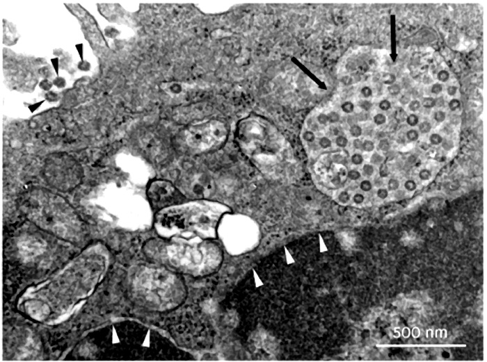
In the case of the novel corona virus, every single published photograph we have seen showing the “isolated” virus shows no such thing. Instead, it shows tissue with a number of dots, usually with an arrow pointing to the so-called coronavirus. If you see tissue in the photograph, by definition, it’s not isolated. An example of such a photograph comes from “Virus Isolation from the First Patient with SARS-CoV-2 in Korea,” published February 24, 2020 in the Journal of Korean Medical Science. Although the authors claim to have isolated the virus, the photographs they publish show “virus” structures inside and outside a cell (indicated by arrows), not isolated.
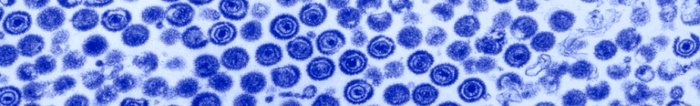
You can see a properly isolated “virus” in the electron microscopy image of the chicken pox “virus,” below. (By the way, although health officials claim that chicken pox is “highly contagious,” no studies have shown that exposing people to isolated chicken pox virus makes them sick.)
What virologists do today is use the liquid—called the supernatant–after either filtration or centrifuging to get rid of the bacteria, fungi and other larger material. This is what they refer to as “purification.” This is like filtering the grounds out of coffee to get caffeine so you can study its effects. But there are hundreds or thousands of other compounds in the coffee, so you still need to isolate the caffeine. What the researchers should then do is put the supernatant in what’s called a sucrose density centrifuge column, which spins out the various compounds into bands. One of these bands will contain the pure virus, which can then be photographed and analyzed.
Instead of working with pure virus, researchers use the supernatant, which contains all kinds of molecules and particles. Instead of doing a genetic analysis of the isolated virus, they do genetic analysis on the mess of compounds in the supernatant.
Now to get enough “virus” to use experimentally, virologists must grow it in a biological medium such as an animal or at least cells from an animal. Unlike bacteria, which can be grown in petri dishes, viruses are not alive and can only “grow” in other living cells.
So they transfer the supernatant not to healthy tissue, but to tissue that has been starved of nutrients and poisoned with strong antibiotics—to make sure that what is left is only viruses and not bacteria and fungi. The main type of tissue they use is kidney cells from various species, often monkey kidney cells (called Vero cells), and lung cancer cells. The “viruses” seem to multiply. The resultant mess of “viruses,” particles, poisons, dead tissue and cellular debris—called “cultured” virus– is then sold to researchers as samples of “purified virus” for them to use in studies.
By the way, the CDC has published guidelines on “transport medium” for viruses. This is what they use to inoculate the starved tissue which then grows the “virus.” The three main ingredients are fetal bovine serum (extracted from still-living fetal calves and preserved with anti-fungals, among other poisons) along with two highly toxic antibiotics, amphotericin (affectionately called ampho-terrible) and gentamicin. This ungodly mixture is then grown on monkey or fetal kidney cells. Interestingly, all doctors know that the main organ affected by gentamicin and ampho-terrible is the kidneys. So you poison the kidney, the kidney breaks down and then the virologist claims that the virus killed the kidney—without performing any controls. Don’t look behind the curtain, folks!
This practice is fraught with obvious problems for proving it is the virus and not the cancer cells or poisoned kidney cells that are causing disease when these viruses get injected into healthy test animals.
Remember that to prove that a specific virus is making humans or animals sick, they need to find the identical virus in many subjects who are sick with the same symptoms—and then make healthy humans or animals sick by exposing them to this virus. But when researchers try to grow the purified virus on healthy cells, they don’t get a lot of viruses; and when they subject healthy tissue, healthy animals or healthy people to these “viruses,” illness does not result—and this is the wily virus that is going to kill us all!
Why do “viruses” multiply in the starved and poisoned kidney or cancer cells? Because when cells are starved or poisoned, they produce exosomes, which are identical in appearance and characteristics to what are called “viruses.” These tiny particles are helpful, not toxic. They do not attack the cells and then multiply; rather, they are produced inside the cell, often in large amounts, when the cells are stressed by poison and starvation.
Viruses and exosomes are indistinguishable, as we learn from a study entitled “The Role of Extracellular Vesicles as Allies of HIV, HCV and SARS viruses,” published in the journal Viruses, May 2020. To quote from the paper, “The remarkable resemblance between EVs [extracellular vesicles, that is, exosomes] and viruses has caused quite a few problems in the studies focused on the analysis of EVs released during viral infection. Nowadays it is an almost impossible mission to separate EVs and viruses by means of canonical vesicle isolation methods, such as differential ultracentrifugation, because they are frequently co-pelleted due to their similar dimensions. To overcome this problem, different studies have proposed the separation of EVs from virus particles by exploiting their different migration velocity in a density gradient or using the presence of specific markers that distinguish viruses from EVs. However, to date, a reliable method that can actually guarantee a complete separation does not exist [emphasis added]. “
In other words, researchers can’t distinguish viruses from exosomes—that’s because they are the same thing and in reality, all viruses are exosomes. Scientists are discovering that all of these “viruses” originate in our own tissues—they don’t attack us from the outside.
With this background, let’s then look at the study, “Koch’s Postulates fulfilled for SARS Virus.” The researchers took unpurified sediment from the snot of sick people, grew that in lung cancer cells until they got a sufficient quantity of cellular material to work with. Then they centrifuged this mess again, not even attempting to purify any virus from the mixture. Finally, they took this unholy mixture of snot sediment, lung cancer cells and who-knows-what-else and injected that into two unfortunate monkeys. They didn’t do a control group by injecting saline into other monkeys or injecting lung cancer cells into monkeys or even injecting the liquid from the centrifuged material into monkeys. They just injected the cellular-debris-laden goop. One of the monkeys got pneumonia, the other got a rash. That, claim the researchers, is the proof that a “coronavirus” can cause disease and that Koch’s postulates have been satisfied.
“The Coronavirus Unveiled,” appearing in the New York Times, gives the impression that researchers are working with a genuine isolated coronavirus. Nevertheless, the article tells us that “In February, as the new coronavirus swept across China and shut down entire cities, . . . the best pictures anyone had managed to take were low-resolution images, in which the virus looked like a barely discernible smudge.”
How did the researchers isolate the virus? They “doused the viruses with chemicals to render them harmless. . .” In other words, they poisoned them. Then they somehow “concentrated the virus-laden fluid from a quart down to a single drop” after which they flash froze the drop. Then in the microscope they saw structures they called viruses.
This is not the proper way to isolate and characterize a virus, either. Proper isolation involves ultrafiltration and centrifuging–not dousing with chemicals and flash freezing–and then performing various physical, biochemical and immunological analyses.
After seeing these particles—most likely helpful exosomes responding to the poisonous chemicals–the researchers state that “its intimately twisted genes commandeer our biochemistry [and] wrenches into our cellular factories, while others build nurseries for making new viruses.” This is highly imaginative horror-movie speculation, not science.
Virologist have three “hosts” they can use in their attempts to prove that viruses cause illness. After “isolating” the virus, they can expose humans to the virus; they can expose animals to the virus; or they can use tissue cultures taken from various animal or human sources and expose the tissue culture to the virus. Leaving aside the fact that they never actually isolate and purify the virus, which they openly admit, let’s assume that the unpurified fluid they are using does contain the relevant virus and therefore should be able to transmit infection. I
In the history of virology, most virologists have decided not to do their experiments on human subjects as this is considered unethical. In the case of the SARS-CoV-2 virus, we know of no published study that used humans as the test subjects.
Virologists also admit that in the case of most viral infections, there are no studies available proving infection in animals. How a virus can infect and kill humans but not animals is left unexplained. Researchers get around this obvious biological conundrum by saying, “there are no animal models on which to test such-and-such a virus.” In other words, “We know that the virus infects and kills humans even though we’ve never tested the virus on humans because that would be unethical. Therefore, we do our tests on animals, even though when we test animals. they don’t get sick, because they are not proper “hosts” for the virus. So, you’ll just have to trust us.”
In the case of SARS CoV-2, we know of two studies that used unpurified “virus” on animal models, one with hamsters and one with mice. In the hamster study, researchers took the unpurified, lung-cancer-grown, centrifuged animal secretions and squirted it down the throats and into the lungs of a group of unfortunate hamsters. Some but not all of the hamsters got pneumonia and some even died. We have no idea what would have happened if they had squirted plain lung cancer cells into the lungs of these hamsters, probably not anything good. Even more perplexing, some of the hamsters didn’t even get sick at all, which certainly doesn’t square with the deadly contagious virus theory.
In the mouse study, researchers infected both transgenic mice and wild (normal) mice with unpurified virus. None of the wild mice exposed to the “virus” got sick. Of the mice genetically programmed to get sick, a statistically insignificant number either lost some fur luster or had an insignificant weight loss. Thus, scientists have not been able to show that the Covid-19 “virus” causes harm to animals.
The purpose of the study was for a group of about twenty virologists to describe the state of the science dealing with the isolation and purification, and the biological characteristics of the new SARS-CoV-2 virus, and to share this information with other scientists for their own research. A thorough and careful reading of this important paper reveals some shocking findings.
First, in the section titled “Whole Genome Sequencing,” we find that rather than having isolated the virus and sequencing the genome from end to end, they “designed 37 pairs of nested PCRs spanning the genome on the basis of the coronavirus reference sequence. . . “ This means they actually looked at a mere thirty-seven primers out of the approximately thirty thousand base pairs claimed to be the genome of an intact virus. They then took these thirty-seven segments and put them into a computer program, which filled in the rest of the genome.
This computer-generation step—called “whole genome sequencing”–constitutes scientific fraud of the highest order. Here is an equivalency: a group of researchers claim to have found a unicorn because they found a piece of a hoof, a hair from a tail, and a sliver of a horn. They then put that information into a computer and program it to re-create the unicorn, claiming that this computer re-creation is the real unicorn. Of course, they have never actually seen a unicorn so could not possibly have examined its genetic makeup to compare their samples with the actual unicorn’s hair, hooves and horn.
The researchers claim they decided which is the real genome of SARS-CoV-2 by “consensus,” sort of like a vote. As different computer programs will come up with different versions of the imaginary “unicorn,” they come together as a group and decide which is the real imaginary unicorn. (By the way, this is how scientists characterized the measles “virus”—by consensus!)
But the real blockbuster finding in this study comes later, a finding so shocking that it’s hard to believe what we are reading. “Therefore, we examined the capacity of SARS-CoV-2 to infect and replicate in several common primate and human cell lines, including human adenocarcinoma cells (A549), human liver cells (HUH 7.0), and human embryonic kidney cells (HEK-293T). In addition to Vero E6 and Vero CCL81 cells . . . Each cell line was inoculated at high multiplicity of infection and examined 24h post-infection.”
This is the third method virologists use to prove infection and pathogenicity — the method they usually rely on—namely, the inoculation of solutions they say contain the virus onto a variety of tissue cultures. As we have pointed out, such inoculation has never been shown to kill (lyse) the tissue, unless the tissue is first poisoned and starved (grown in a “minimal-nutrient medium.”)
In the Results section, the authors state: “Therefore, we examined the capacity of SARS-CoV-2 to infect and replicate in several common primate and human cell lines, including human adenocarcinoma cells (A549), human liver cells (HUH7.0), and human embryonic kidney cells (HEK-293T) . . . Each cell line was inoculated at high multiplicity of infection and examined 24h post infection. No CPE was observed in any of the cell lines except in Vero cells.”
Note, CPE means “cytopathic effect,” which refers to structural changes in host cells that are caused by “viral invasion.” The infecting virus is said to cause lysis (breaking up) of the host cell or, when the cell dies without lysis, an inability to reproduce. Both of these effects are said to occur due to CPEs.
So did this viral material with its “intimately twisted genes commandeer the cellular biochemistry [and] wrench into the cellular factories, while others build nurseries for making new viruses?” Nothing of the sort!
The shocking thing about the findings is that using their own methods, the virologists found that solutions claimed to contain SARS-CoV-2 (as well as poisons)—even in high amounts –were not infective to any of the three human tissue cultures they tested. In plain English, this means they proved, on their terms, that this “new coronavirus” is not infectious to human beings. It is only infective to monkey kidney cells, and only when you add two potent drugs (gentamicin and amphotericin), known to be toxic to kidneys, to the mix.
Interestingly, in their conclusion the authors don’t mention this important fact. Only virologists reading the whole paper will find out that if you want to grow the virus, don’t bother to use human cell lines.
Meanwhile we have worldwide lockdown predicated on the idea that something called coronavirus causes disease. As you can read, in all three of the human cell lines no CPE (no cell death, no infection) was observed. Only Vero cells (monkey kidney cells) were adversely affected—and remember, the material injected into these cells contained kidney toxins. So basically, they proved that the SARS-CoV-2 virus does not infect human tissue.
The researchers used “The wild type chimpanzee adenovirus isolate Y25 [which] was originally obtained from William Hillis, John Hopkins University of Medicine. The virus was passaged in HEK293A cells (Invitrogen, Cat. R705-07) and purified by CsCl gradient . . . Viral DNA was phenol extracted for genomic sequencing and cloning.”
The researchers purchased some material (not properly isolated even though it is called an “isolate”) which they then “passaged” through human embryonic kidney cells (called HEK293A), and then they “purified” it by CsCl gradient. You can read about this technique here. It separates DNA molecules (not viruses) after mixing them with cesium chloride (a heavy metal salt) and ethidium bromide (a mutagen that can affect DNA biological processes, like DNA replication and transcription.)
This is the same smoke and mirrors—not true separation and isolation but “surrogate” techniques that use various poisons.
Another study sent to us is entitled, “SARS-CoV-2 structure and replication characterized by in situ cryo-electron tomography,” published June 23, 2020. The authors begin with the creed of the faithful: “β-coronaviruses, including SARS-CoV-1 and Middle Eastern Respiratory Virus (MERS-CoV) are highly contagious pathogens that can cause severe lower respiratory infections. At the end of 2019, SARS-CoV-2 emerged in the city of Wuhan, China, likely through zoonotic transmission via a bat reservoir and a still unidentified intermediate host that subsequently led to a pandemic, accumulating to date to over 8 million cases and close to 500,000 deaths worldwide.”
The article provides no references for the statement that the SARS virus is “highly contagious” but does contain a lot of fuzzy electron-microscope photographs of tissues and cells whose genetic material they determined using PCR tests—the equivalent of finding moats and turrets in a bunch of Lego pieces.
The researchers did not isolate and purify the virus but instead used “monkey kidney derived VeroE6 cells” and “human pulmonary cell lines.” In other words, they used cell lines grown in starved and poisoned cultures.
Later in the paper the authors state that they get different “morphologies” of the virus depending on which cell line they use. In other words when grown on monkey kidney cells the virus looks one way, grown on lung cancer the same virus looks different. That is like saying that if you plant some seeds in one garden you will get tomatoes but if you plant them in another garden you will get turnips. What this observation tells us is that what they find comes from the tissue not the source “virus,” that is why they are different.
According to the authors, “Our report provides the first in situ cryo-ET analysis of coronaviruses at high preservation levels.” Wait a minute—this study was published on June 23, 2020. You mean they had no analyses of this virus before health officials called for universal lockdown?
Says Scoglio, “At the center of the pandemic project stands the Covid swab test, which is based on the RT-PCR (Reverse Transcriptase- Polymerase Chain reaction): a sample of organic material is taken from the throat, or more rarely from the broncho-alveolar fluid, of the individual, and then the presence of the SARS-Cov-2 virus in the sample is tested. This is done by using the same RT-PCR methodology used to originally “isolate” the virus from patient zero. Thus, the Covid test depends essentially on the original isolation, or lack thereof, of the SARS-Cov2 virus, the original PCR isolation of the virus constituting the golden standard necessary to validate any subsequent Covid test. The problems with the original virus isolation, and thus with the ensuing swab test, are many, and they all point to the truth that the SARS-Cov2 virus has never been isolated and never tested for its pathogenicity.”
One argument we hear is that Koch’s postulates are irrelevant, out of date, useless or even “wrong.” If so, why do researchers claim to have satisfied Koch’s postulates, not only for Covid-19 but for other diseases like HIV/AIDS and Lyme’s disease.
For example, in 1997, scientists announced that human immunodeficiency virus (HIV) does fulfill Koch’ postulates and hence is the proven cause of AIDS. The study involved taking the blood from an HIV-positive person and injecting it into one chimpanzee. They didn’t purify or isolate anything, just injected the blood into one chimpanzee. They kept the chimp for ten years–who knows what they fed it or anything about its conditions of confinement. After ten years the chimp developed an “opportunistic infection” (which could even be a yeast infection) and tested HIV-positive (a test result that occurs in at least thirty-three other conditions). The study had no controls–like injecting the chimp with blood from someone with cancer or with blood from a healthy person. This was the proof that HIV causes AIDS! This is not science, but it keeps the grant money flowing.
With Lyme’s disease the “proof” that Koch’s postulates were fulfilled comes from a paper published in 1983, which reported detection of spirochete [spiral-shaped bacteria] in the blood of two patients with Lyme. The researchers then examined some ticks in the neighborhood and found the same spirochete. That’s it, that was the “proof” of Koch’s postulates.
As we have explained, finding bacteria at the site of an injury or in a person with a disease in no way constitutes proof of causation any more than finding firemen at the site of a fire means they caused the fire. Among other roles, bacteria act as scavengers in nature, they “eat” dead or diseased tissue. Maggots play the same role; if you see a dead dog crawling with maggots, it would be crazy conclude that the maggots killed the dog. So why do scientists assume that the presence of “viruses” in a cell means that the cell has been attacked from the outside and taken over by hostile compounds?
If anyone can show us a properly done study in which the “coronavirus” from many sick people was isolated, purified, photographed and characterized according to the consensus agreement of the 1973 Pasteur Institute guidelines, and then shown to cause disease in healthy organisms (animals or humans), we will gladly withdraw the book. Meanwhile, we contend that the idea of a contagious coronavirus is a fairy tale.
The Contagion Myth is banned on Amazon but available at:
 Few of us were born when the forces for milk pasteurization launched the first major attack on Nature’s perfect food. In 1945, a magazine called Coronet published an article, “Raw Milk Can Kill You,” blaming raw milk for an outbreak of brucellosis in a town called Crossroads, U.S.A., killing one-third of the inhabitants. The Reader’s Digest picked up the story and ran it a year later.