The Contagion Fairy Tale
by Thomas Cowan, MD and Sally Fallon Morell
November 10, 2020
Our book, The Contagion Myth, is now available (banned on Amazon but sold on other outlets) and has already generated dozens of comments, many of them challenging our contention that the corona “virus” does not exist and that the illness attributed to this virus is not contagious—one referred to our book as a fairy tale!

However, unlike most coronavirus skeptics, we are not arguing that the illness is just a bad case of the flu, with deaths due solely to pre-existing conditions or inappropriate hospital care; rather we postulate that the illness can be very serious and that the likely cause is radiation poisoning, probably from the worldwide deployment of 5G, starting in Wuhan, China and followed by major cities throughout the world.
Comments we have received include the following:
- Okinawa does not have 5G but people are getting infected there;
- Some friends went to a wedding in Kirkland, Washington and got Covid, so it must be infectious;
- There’s 5G in New Zealand but very few cases of illness;
- A school in our neighborhood has opened for in-person classes and there has been an outbreak—two people have tested positive;
- A lot of people “got the virus” after a big no-mask motorcycle rally in Sturgis, South Dakota;
- What about rabbits getting myxomatosis, a known viral disease.
With the exception of the rabbit comment (a subject to be explored in a future blog), these observations are just that—epidemiological observations, which are certainly interesting and deserve further exploration, but these in no way disprove our main contention that this virus does not exist and the illness attributed to it is not contagious.
Why take our word for the shocking claim that no scientist has found the so-called coronavirus? Of course, you shouldn’t take our word for it, you should listen to what the experts are saying. In July 2020, the FDA posted a CDC document entitled “CDC 2019-Novel Coronavirus (2019-nCoV), Real-Time RT-PCR diagnostic Panel. For Emergency Use Only. Instructions for Use.” Buried in the text, on page 39, is the following statement: “. . . no quantified virus isolates of the 2019-nCoV are currently available.”
In other words, our government is telling us that there are no purified isolated samples of this “novel coronavirus,” which means that the virus has never been isolated and purified. What they are finding in the RT-PCR tests are fragments of genetic material, which actually come from human chromosome #8. This means that the results of all RT-PCR tests are invalid—the only thing they can tell us is that we are human beings.
A January, 2020 paper on testing tells us the same thing: “The ongoing outbreak of the recently emerged novel coronavirus (2019-nCoV) poses a challenge for public health laboratories as virus isolates are unavailable. . . [emphasis added]” Nevertheless, even without knowing what this virus is like, the researchers aim “to develop and deploy robust diagnostic methodology for use in public health laboratory settings without having virus material available.” A challenge indeed!
Here is an analogy to describe what is going on. Let’s say you are a paid Lego specialist and someone offers to reward you if you can construct an exact replica of King Beauregard’s Medieval castle. The referees put all the known Lego pieces out on a table and promise to pay you well to do the reconstruction. Naturally, you ask to see a picture of what the castle looked like or at least some sort of architectural plan so you know what to build. But the referees say that you must reconstruct the castle without having access to any information about the original castle. You think this is downright bizarre, but since a job is a job, you start looking. You find pieces for a moat; you know that castles have moats and think that this must be part of the castle. Then you find windows, turrets, soldiers, etc.–with each new finding you are given a castle-building Lego award and an increase in salary. You write some software that fills in the rest of the castle from the fragments you have. Then you publish a peer reviewed paper on the “completed” castle for all the world to see.
Unfortunately, a child appears who looks like he has time traveled from the Middle Ages. You show him the castle. “Everybody knows that Beauregard didn’t have a castle.” He says. “Beauregard was an impoverished aristocrat who was afraid of moats; he lived in a garret in London.” But the show must go on, so his remarks are never published, while the Lego expert (who knows the child is right) keeps quiet and enjoys his hefty salary.
A number of readers have sent us studies “proving” the existence of pathogenic viruses. In fact, one virologist claimed that “thousands of papers” show that isolated bacteria or viruses cause disease. (He also tried to convince us that one could sterilize one’s hands, cover them and they would remain sterile “indefinitely.”)
One was a link to a study with the promising title “Koch’s postulates fulfilled for SARS virus”, published 2003 in the prestigious journal Nature. We discuss this study in The Contagion Myth. The researchers claim that Severe Acute Respiratory Syndrome (SARS) is also caused by a coronavirus. The title itself is misleading, not to say fraudulent, because the researchers did not satisfy Koch’s postulates—which is the common-sense way of proving that a microbe causes disease. They did not satisfy River’s postulates either—River’s postulates are for proving that a virus causes a disease. These methods involve isolating and purifying a specific microbial organism from a number of individuals suffering from a specific disease and injecting the isolated, purified bacteria or virus into healthy organisms (animal or human). If every sick person has the organism and every test subject becomes ill, then you know that the specific microbe causes the specific disease.
Let’s focus on the process of isolating and purifying a virus—it’s hard to do but not impossible. In 1973, the Pasteur Institute published guidelines for doing this. First the virologist takes mucus or secretions from a person with the disease. The secretions are diluted and then put into a kind of blender. The resultant liquid is then passed through a very fine filter—fine enough to keep out bacteria and fungi but let the viruses through; the resulting liquid is called a supernatant. It contains the virus but also lots of other stuff as well. The supernatant must then be centrifuged in such a way that you get bands of particles of the same size and weight. The scientist can determine which band is the virus using the known size and weight of viruses. This band is removed from the supernatant with a pipette. This is the properly isolated and purified virus. The virus is then transferred to some tissue to grow and multiply.
An important point is that when the virologist has finished the purification process of macerating, filtering and ultracentrifugation, he must then take an electron micrograph of the final, purified virus to show his colleagues that he has in fact successfully purified and isolated the virus. Virologists have done this many times and for many different viruses. Without an electron micrograph picture showing purification, no reputable journal would publish this work. The reason is simple: scientists are essentially told not to believe each other because someone says so. If you say you isolated a virus you must show the picture to prove it, period. Absent the picture it could be a total fabrication. The way science is supposed to work, after you have isolated and photographed the virus, other scientists in other labs follow the exact steps that you outlined in your paper and show pictures of the same isolated virus. Once a number of labs have done this, you have real proof that the virus exists.
In the case of the novel corona virus, every single published photograph we have seen showing the “isolated” virus shows no such thing. Instead, it shows tissue with a number of dots, usually with an arrow pointing to the so-called coronavirus. If you see tissue in the photograph, by definition, it’s not isolated. An example of such a photograph comes from “Virus Isolation from the First Patient with SARS-CoV-2 in Korea,” published February 24, 2020 in the Journal of Korean Medical Science. Although the authors claim to have isolated the virus, the photographs they publish show “virus” structures inside and outside a cell (indicated by arrows), not isolated.
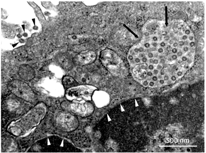
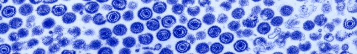
You can see a properly isolated “virus” in the electron microscopy image of the chicken pox “virus,” below. (By the way, although health officials claim that chicken pox is “highly contagious,” no studies have shown that exposing people to isolated chicken pox virus makes them sick.)
What virologists do today is use the liquid—called the supernatant–after either filtration or centrifuging to get rid of the bacteria, fungi and other larger material. This is what they refer to as “purification.” This is like filtering the grounds out of coffee to get caffeine so you can study its effects. But there are hundreds or thousands of other compounds in the coffee, so you still need to isolate the caffeine. What the researchers should then do is put the supernatant in what’s called a sucrose density centrifuge column, which spins out the various compounds into bands. One of these bands will contain the pure virus, which can then be photographed and analyzed.
Instead of working with pure virus, researchers use the supernatant, which contains all kinds of molecules and particles. Instead of doing a genetic analysis of the isolated virus, they do genetic analysis on the mess of compounds in the supernatant.
Now to get enough “virus” to use experimentally, virologists must grow it in a biological medium such as an animal or at least cells from an animal. Unlike bacteria, which can be grown in petri dishes, viruses are not alive and can only “grow” in other living cells.
So they transfer the supernatant not to healthy tissue, but to tissue that has been starved of nutrients and poisoned with strong antibiotics—to make sure that what is left is only viruses and not bacteria and fungi. The main type of tissue they use is kidney cells from various species, often monkey kidney cells (called Vero cells), and lung cancer cells. The “viruses” seem to multiply. The resultant mess of “viruses,” particles, poisons, dead tissue and cellular debris—called “cultured” virus– is then sold to researchers as samples of “purified virus” for them to use in studies.
By the way, the CDC has published guidelines on “transport medium” for viruses. This is what they use to inoculate the starved tissue which then grows the “virus.” The three main ingredients are fetal bovine serum (extracted from still-living fetal calves and preserved with anti-fungals, among other poisons) along with two highly toxic antibiotics, amphotericin (affectionately called ampho-terrible) and gentamicin. This ungodly mixture is then grown on monkey or fetal kidney cells. Interestingly, all doctors know that the main organ affected by gentamicin and ampho-terrible is the kidneys. So you poison the kidney, the kidney breaks down and then the virologist claims that the virus killed the kidney—without performing any controls. Don’t look behind the curtain, folks!
This practice is fraught with obvious problems for proving it is the virus and not the cancer cells or poisoned kidney cells that are causing disease when these viruses get injected into healthy test animals.
Remember that to prove that a specific virus is making humans or animals sick, they need to find the identical virus in many subjects who are sick with the same symptoms—and then make healthy humans or animals sick by exposing them to this virus. But when researchers try to grow the purified virus on healthy cells, they don’t get a lot of viruses; and when they subject healthy tissue, healthy animals or healthy people to these “viruses,” illness does not result—and this is the wily virus that is going to kill us all!
Why do “viruses” multiply in the starved and poisoned kidney or cancer cells? Because when cells are starved or poisoned, they produce exosomes, which are identical in appearance and characteristics to what are called “viruses.” These tiny particles are helpful, not toxic. They do not attack the cells and then multiply; rather, they are produced inside the cell, often in large amounts, when the cells are stressed by poison and starvation.
Viruses and exosomes are indistinguishable, as we learn from a study entitled “The Role of Extracellular Vesicles as Allies of HIV, HCV and SARS viruses,” published in the journal Viruses, May 2020. To quote from the paper, “The remarkable resemblance between EVs [extracellular vesicles, that is, exosomes] and viruses has caused quite a few problems in the studies focused on the analysis of EVs released during viral infection. Nowadays it is an almost impossible mission to separate EVs and viruses by means of canonical vesicle isolation methods, such as differential ultracentrifugation, because they are frequently co-pelleted due to their similar dimensions. To overcome this problem, different studies have proposed the separation of EVs from virus particles by exploiting their different migration velocity in a density gradient or using the presence of specific markers that distinguish viruses from EVs. However, to date, a reliable method that can actually guarantee a complete separation does not exist [emphasis added]. “
In other words, researchers can’t distinguish viruses from exosomes—that’s because they are the same thing and in reality, all viruses are exosomes. Scientists are discovering that all of these “viruses” originate in our own tissues—they don’t attack us from the outside.
With this background, let’s then look at the study, “Koch’s Postulates fulfilled for SARS Virus.” The researchers took unpurified sediment from the snot of sick people, grew that in lung cancer cells until they got a sufficient quantity of cellular material to work with. Then they centrifuged this mess again, not even attempting to purify any virus from the mixture. Finally, they took this unholy mixture of snot sediment, lung cancer cells and who-knows-what-else and injected that into two unfortunate monkeys. They didn’t do a control group by injecting saline into other monkeys or injecting lung cancer cells into monkeys or even injecting the liquid from the centrifuged material into monkeys. They just injected the cellular-debris-laden goop. One of the monkeys got pneumonia, the other got a rash. That, claim the researchers, is the proof that a “coronavirus” can cause disease and that Koch’s postulates have been satisfied.
“The Coronavirus Unveiled,” appearing in the New York Times, gives the impression that researchers are working with a genuine isolated coronavirus. Nevertheless, the article tells us that “In February, as the new coronavirus swept across China and shut down entire cities, . . . the best pictures anyone had managed to take were low-resolution images, in which the virus looked like a barely discernible smudge.”
How did the researchers isolate the virus? They “doused the viruses with chemicals to render them harmless. . .” In other words, they poisoned them. Then they somehow “concentrated the virus-laden fluid from a quart down to a single drop” after which they flash froze the drop. Then in the microscope they saw structures they called viruses.
This is not the proper way to isolate and characterize a virus, either. Proper isolation involves ultrafiltration and centrifuging–not dousing with chemicals and flash freezing–and then performing various physical, biochemical and immunological analyses.
After seeing these particles—most likely helpful exosomes responding to the poisonous chemicals–the researchers state that “its intimately twisted genes commandeer our biochemistry [and] wrenches into our cellular factories, while others build nurseries for making new viruses.” This is highly imaginative horror-movie speculation, not science.
Virologist have three “hosts” they can use in their attempts to prove that viruses cause illness. After “isolating” the virus, they can expose humans to the virus; they can expose animals to the virus; or they can use tissue cultures taken from various animal or human sources and expose the tissue culture to the virus. Leaving aside the fact that they never actually isolate and purify the virus, which they openly admit, let’s assume that the unpurified fluid they are using does contain the relevant virus and therefore should be able to transmit infection. I
In the history of virology, most virologists have decided not to do their experiments on human subjects as this is considered unethical. In the case of the SARS-CoV-2 virus, we know of no published study that used humans as the test subjects.
Virologists also admit that in the case of most viral infections, there are no studies available proving infection in animals. How a virus can infect and kill humans but not animals is left unexplained. Researchers get around this obvious biological conundrum by saying, “there are no animal models on which to test such-and-such a virus.” In other words, “We know that the virus infects and kills humans even though we’ve never tested the virus on humans because that would be unethical. Therefore, we do our tests on animals, even though when we test animals. they don’t get sick, because they are not proper “hosts” for the virus. So, you’ll just have to trust us.”
In the case of SARS CoV-2, we know of two studies that used unpurified “virus” on animal models, one with hamsters and one with mice. In the hamster study, researchers took the unpurified, lung-cancer-grown, centrifuged animal secretions and squirted it down the throats and into the lungs of a group of unfortunate hamsters. Some but not all of the hamsters got pneumonia and some even died. We have no idea what would have happened if they had squirted plain lung cancer cells into the lungs of these hamsters, probably not anything good. Even more perplexing, some of the hamsters didn’t even get sick at all, which certainly doesn’t square with the deadly contagious virus theory.
In the mouse study, researchers infected both transgenic mice and wild (normal) mice with unpurified virus. None of the wild mice exposed to the “virus” got sick. Of the mice genetically programmed to get sick, a statistically insignificant number either lost some fur luster or had an insignificant weight loss. Thus, scientists have not been able to show that the Covid-19 “virus” causes harm to animals.
The third choice for virologist is to infect human and animal tissue with a “culture” of the virus to see what happens. This is what they did in a study entitled, “Severe Acute Respiratory Syndrome Coronavirus 2 from Patient with Coronavirus Disease,” published in the CDC Bulletin, June 2020.
The purpose of the study was for a group of about twenty virologists to describe the state of the science dealing with the isolation and purification, and the biological characteristics of the new SARS-CoV-2 virus, and to share this information with other scientists for their own research. A thorough and careful reading of this important paper reveals some shocking findings.
First, in the section titled “Whole Genome Sequencing,” we find that rather than having isolated the virus and sequencing the genome from end to end, they “designed 37 pairs of nested PCRs spanning the genome on the basis of the coronavirus reference sequence. . . “ This means they actually looked at a mere thirty-seven primers out of the approximately thirty thousand base pairs claimed to be the genome of an intact virus. They then took these thirty-seven segments and put them into a computer program, which filled in the rest of the genome.
This computer-generation step—called “whole genome sequencing”–constitutes scientific fraud of the highest order. Here is an equivalency: a group of researchers claim to have found a unicorn because they found a piece of a hoof, a hair from a tail, and a sliver of a horn. They then put that information into a computer and program it to re-create the unicorn, claiming that this computer re-creation is the real unicorn. Of course, they have never actually seen a unicorn so could not possibly have examined its genetic makeup to compare their samples with the actual unicorn’s hair, hooves and horn.
The researchers claim they decided which is the real genome of SARS-CoV-2 by “consensus,” sort of like a vote. As different computer programs will come up with different versions of the imaginary “unicorn,” they come together as a group and decide which is the real imaginary unicorn. (By the way, this is how scientists characterized the measles “virus”—by consensus!)
But the real blockbuster finding in this study comes later, a finding so shocking that it’s hard to believe what we are reading. “Therefore, we examined the capacity of SARS-CoV-2 to infect and replicate in several common primate and human cell lines, including human adenocarcinoma cells (A549), human liver cells (HUH 7.0), and human embryonic kidney cells (HEK-293T). In addition to Vero E6 and Vero CCL81 cells . . . Each cell line was inoculated at high multiplicity of infection and examined 24h post-infection.”
This is the third method virologists use to prove infection and pathogenicity — the method they usually rely on—namely, the inoculation of solutions they say contain the virus onto a variety of tissue cultures. As we have pointed out, such inoculation has never been shown to kill (lyse) the tissue, unless the tissue is first poisoned and starved (grown in a “minimal-nutrient medium.”)
In the Results section, the authors state: “Therefore, we examined the capacity of SARS-CoV-2 to infect and replicate in several common primate and human cell lines, including human adenocarcinoma cells (A549), human liver cells (HUH7.0), and human embryonic kidney cells (HEK-293T) . . . Each cell line was inoculated at high multiplicity of infection and examined 24h post infection. No CPE was observed in any of the cell lines except in Vero cells.”
Note, CPE means “cytopathic effect,” which refers to structural changes in host cells that are caused by “viral invasion.” The infecting virus is said to cause lysis (breaking up) of the host cell or, when the cell dies without lysis, an inability to reproduce. Both of these effects are said to occur due to CPEs.
So did this viral material with its “intimately twisted genes commandeer the cellular biochemistry [and] wrench into the cellular factories, while others build nurseries for making new viruses?” Nothing of the sort!
The shocking thing about the findings is that using their own methods, the virologists found that solutions claimed to contain SARS-CoV-2 (as well as poisons)—even in high amounts –were not infective to any of the three human tissue cultures they tested. In plain English, this means they proved, on their terms, that this “new coronavirus” is not infectious to human beings. It is only infective to monkey kidney cells, and only when you add two potent drugs (gentamicin and amphotericin), known to be toxic to kidneys, to the mix.
Interestingly, in their conclusion the authors don’t mention this important fact. Only virologists reading the whole paper will find out that if you want to grow the virus, don’t bother to use human cell lines.
Meanwhile we have worldwide lockdown predicated on the idea that something called coronavirus causes disease. As you can read, in all three of the human cell lines no CPE (no cell death, no infection) was observed. Only Vero cells (monkey kidney cells) were adversely affected—and remember, the material injected into these cells contained kidney toxins. So basically, they proved that the SARS-CoV-2 virus does not infect human tissue.
Another study sent to us comes with the fancy title, “A Novel Chimpanzee Adenovirus Vector with Low Human Seroprevalence: Improved Systems for Vector Derivation and Comparative Immunogenicity.”
The researchers used “The wild type chimpanzee adenovirus isolate Y25 [which] was originally obtained from William Hillis, John Hopkins University of Medicine. The virus was passaged in HEK293A cells (Invitrogen, Cat. R705-07) and purified by CsCl gradient . . . Viral DNA was phenol extracted for genomic sequencing and cloning.”
The researchers purchased some material (not properly isolated even though it is called an “isolate”) which they then “passaged” through human embryonic kidney cells (called HEK293A), and then they “purified” it by CsCl gradient. You can read about this technique here. It separates DNA molecules (not viruses) after mixing them with cesium chloride (a heavy metal salt) and ethidium bromide (a mutagen that can affect DNA biological processes, like DNA replication and transcription.)
This is the same smoke and mirrors—not true separation and isolation but “surrogate” techniques that use various poisons.
Another study sent to us is entitled, “SARS-CoV-2 structure and replication characterized by in situ cryo-electron tomography,” published June 23, 2020. The authors begin with the creed of the faithful: “β-coronaviruses, including SARS-CoV-1 and Middle Eastern Respiratory Virus (MERS-CoV) are highly contagious pathogens that can cause severe lower respiratory infections. At the end of 2019, SARS-CoV-2 emerged in the city of Wuhan, China, likely through zoonotic transmission via a bat reservoir and a still unidentified intermediate host that subsequently led to a pandemic, accumulating to date to over 8 million cases and close to 500,000 deaths worldwide.”
The article provides no references for the statement that the SARS virus is “highly contagious” but does contain a lot of fuzzy electron-microscope photographs of tissues and cells whose genetic material they determined using PCR tests—the equivalent of finding moats and turrets in a bunch of Lego pieces.
The researchers did not isolate and purify the virus but instead used “monkey kidney derived VeroE6 cells” and “human pulmonary cell lines.” In other words, they used cell lines grown in starved and poisoned cultures.
Later in the paper the authors state that they get different “morphologies” of the virus depending on which cell line they use. In other words when grown on monkey kidney cells the virus looks one way, grown on lung cancer the same virus looks different. That is like saying that if you plant some seeds in one garden you will get tomatoes but if you plant them in another garden you will get turnips. What this observation tells us is that what they find comes from the tissue not the source “virus,” that is why they are different.
According to the authors, “Our report provides the first in situ cryo-ET analysis of coronaviruses at high preservation levels.” Wait a minute—this study was published on June 23, 2020. You mean they had no analyses of this virus before health officials called for universal lockdown?
By the way, Stefano Scoglio, PhD, from Italy, has come to the same conclusions that we have in a talk entitled “THE INVENTED PANDEMIC, the lack of VIRUS ISOLATION and the INVALID COVID-19 test.”
Says Scoglio, “At the center of the pandemic project stands the Covid swab test, which is based on the RT-PCR (Reverse Transcriptase- Polymerase Chain reaction): a sample of organic material is taken from the throat, or more rarely from the broncho-alveolar fluid, of the individual, and then the presence of the SARS-Cov-2 virus in the sample is tested. This is done by using the same RT-PCR methodology used to originally “isolate” the virus from patient zero. Thus, the Covid test depends essentially on the original isolation, or lack thereof, of the SARS-Cov2 virus, the original PCR isolation of the virus constituting the golden standard necessary to validate any subsequent Covid test. The problems with the original virus isolation, and thus with the ensuing swab test, are many, and they all point to the truth that the SARS-Cov2 virus has never been isolated and never tested for its pathogenicity.”
One argument we hear is that Koch’s postulates are irrelevant, out of date, useless or even “wrong.” If so, why do researchers claim to have satisfied Koch’s postulates, not only for Covid-19 but for other diseases like HIV/AIDS and Lyme’s disease.
For example, in 1997, scientists announced that human immunodeficiency virus (HIV) does fulfill Koch’ postulates and hence is the proven cause of AIDS. The study involved taking the blood from an HIV-positive person and injecting it into one chimpanzee. They didn’t purify or isolate anything, just injected the blood into one chimpanzee. They kept the chimp for ten years–who knows what they fed it or anything about its conditions of confinement. After ten years the chimp developed an “opportunistic infection” (which could even be a yeast infection) and tested HIV-positive (a test result that occurs in at least thirty-three other conditions). The study had no controls–like injecting the chimp with blood from someone with cancer or with blood from a healthy person. This was the proof that HIV causes AIDS! This is not science, but it keeps the grant money flowing.
With Lyme’s disease the “proof” that Koch’s postulates were fulfilled comes from a paper published in 1983, which reported detection of spirochete [spiral-shaped bacteria] in the blood of two patients with Lyme. The researchers then examined some ticks in the neighborhood and found the same spirochete. That’s it, that was the “proof” of Koch’s postulates.
As we have explained, finding bacteria at the site of an injury or in a person with a disease in no way constitutes proof of causation any more than finding firemen at the site of a fire means they caused the fire. Among other roles, bacteria act as scavengers in nature, they “eat” dead or diseased tissue. Maggots play the same role; if you see a dead dog crawling with maggots, it would be crazy conclude that the maggots killed the dog. So why do scientists assume that the presence of “viruses” in a cell means that the cell has been attacked from the outside and taken over by hostile compounds?
If anyone can show us a properly done study in which the “coronavirus” from many sick people was isolated, purified, photographed and characterized according to the consensus agreement of the 1973 Pasteur Institute guidelines, and then shown to cause disease in healthy organisms (animals or humans), we will gladly withdraw the book. Meanwhile, we contend that the idea of a contagious coronavirus is a fairy tale.
The Contagion Myth is banned on Amazon but available at:
Dr. Cowan’s website https://drtomcowan.com/products/the-contagion-myth/


Truth Comes to Light highlights writers and video creators who ask the difficult questions while sharing their unique insights and visions.
Everything posted on this site is done in the spirit of conversation. Please do your own research and trust yourself when reading and giving consideration to anything that appears here or anywhere else.