Inventing the Nature of “Viruses”


“Why is it so difficult for virologists to simply explain basic questions about a ‘virus’ such as whether the ‘virus’ is living or dead? Why must the concept of what a ‘virus’ is change depending upon the researchers and technology of the time? What physical organism changes in concept after over a century of supposed study? The answer to all of these questions is actually fairly easy to grasp. As the researchers have never actually had any physical entities on hand in order to study, the concept of what the assumed invisible entities are was allowed to constantly change in order to suit the needs and evidence of the researchers of the time. There was no solid foundation for virology to stand upon from the very beginning in order to definitively state what the nature of a ‘virus’ truly is.”


Inventing the Nature of “Viruses”

by Mike Stone, Viroliegy
November 10, 2023

“No viruses have been found multiplying free in nature.”

-Virologist Thomas Rivers
Tom Rivers: reflections on a life in medicine and science : an oral history memoir

For the greater part of the first 50 years of the 20th century, there was no agreed upon definition for what the invisible entities labelled as a “virus” actually were nor how these agents looked, formed and functioned. Some researchers believed that these entities were endogenous processes produced within the host while others envisioned them as exogenous invaders that came from outside and attacked from within. There were arguments over whether “viruses” were corpuscular in nature or whether they were a soluble liquid. Debates centered around whether these agents were alive or if they were simply inanimate and non-living. While there were researchers who believed “viruses” were a ferment or a chemical molecule of some kind, the majority believed that these invisible entities were just smaller unseen bacterium. According to biochemist and historian of science Ton van Helvoort’s 1996 paper When Did Virology Start?, the “virus” concept lacked clarity and certainty over the first half of the 20th century. However, the link between bacteriology and “viruses” was so strong at this time that these unseen entities were not considered conceptually distinct from bacteria:

“I have come to believe that, despite its widespread appearance in textbooks and journals of that era, the early concept of the “filterable virus” lacked clarity and certainty. More importantly, I also believe that during the 1930s and 194Os, the links between the study of filterable viruses and bacteriology were so strong that viruses were still considered merely another form of bacteria-not conceptually distinct, as they now are.”

The reason for these many contradictory ideas about the nature of the “virus” was a direct result of the fact that the researchers never had a physical entity on hand in order to study. The “virus” was nothing more than a fluid concept that was open to the interpretation of those who claimed to be working with them. Most of these researchers came from a bacteriological or chemistry background, and thus, they viewed the “virus” concept through their own lens and paradigms. Regardless, there was no way to actually determine the true nature of something that could not be seen or studied in reality and that only existed within the realm of the imagination.

Thus, it shouldn’t be hard to understand why virologists often have a difficult time answering simple questions such as “What is a virus?” or “Is it alive or dead?” This is exactly the argument made in the appropriately titled 2014 article Inventing Viruses by William Summers, a retired Professor of Therapeutic Radiology, Molecular Biophysics & Biochemistry, and History of Medicine. While being able to define what a “virus” is should be an easy task for any virologist, simple questions about the nature of a “virus” are not ones that are simple for them to answer. In the opening of his paper, Summers asked a more subtle question about the invention of the “virus” category:

“…how generations of microbiologists arrived at the idea that some of the entities they dealt with fell into a category that differed in fundamental ways from others. In other words, how did they invent the category of “virus” as we now know it?”

Summers looked to investigate how the idea that “viruses” are a separate entity that requires its own category away from bacteriology came to be. In doing so, he admitted that our beliefs, understandings, and conceptions of what a “virus” is changes over time. This is because “viruses” are whatever a virologist tells us that they are. The concept and the nature of the “virus” was invented, and continually reinvented, by virologists as part of the normal progress of their (pseudo) science. In other words, the idea of the “virus” is able to change at any time based upon whatever a virologist wants a “virus” to be at any given moment:

“Even so, how did the category “virus” come to be recognized, and what are its essential, defining qualities? Viruses are natural objects, but our beliefs, understanding, and conceptions of them change over time on the basis of new information, new points of view, and new scientific values and standards. In a very real way, a virus is what virologists say it is. It is a product of the way virologists talk about viruses—that is, the way facts about viruses are organized in their discourse. It can be said that virologists invent (and continually reinvent) the concept of a virus as part of the normal progress of their science.”

The deliberate ever-changing concept of the “virus” shifted away from its original invention as an agent of disease transmission to its modern day concept as a genetic assembly that sometimes causes disease when it integrates into its host in order to survive. This reinvention of the concept happened in 1957 when French microbiologist Andre Lwoff took many competing and contradictory ideas and mashed them together into the modern definition of a “virus” based upon work done with bacteriophages. Prior to his reinvention of the concept, in 1953, Lwoff actually questioned whether a bacteriophage was a “virus” and wanted to know exactly what a “virus” was. He even noted that “viruses” are defined to be exogenous (coming from outside of the body) while bacteriophages are “always formed inside its host” and “could therefore be described as endogenous,” i.e. originating from within the host. In fact, Lwoff stated that “if prophage is phylogenetically endogenous, the temperate phage produced by a lysogenic bacterium must be described as endogenous,” meaning that the phage is from within the host, thus negating it as an exogenous entity in line with the definition of a “virus.” Ironically, after redefining the “virus” as a genetic code in 1957, Lwoff would ultimately warn in 1991 that virology was “in danger of losing its soul, since viruses now show a strong tendency to become sequences.” He also argued that the abundance of discoveries was causing “the very concept of virus” to waver “on its foundations,” noting that the “problem today and in future is to keep abreast of its whereabouts.”

Regardless, Summers stated that his paper was not about the “triumphant accumulation of knowledge by the heroic scientists” of the past. Rather, it was an examination of the “continual struggle to understand and organize observations.” This struggle was showcased by Lwoff’s own attempts to rationalize and combine contradictory evidence in order to create the modern genetic concept of the “virus” from an entity that did not meet the necessary requirements:

“Nobelist Andre Lwoff, perhaps in a Gertrude Stein frame of mind, famously answered “viruses are viruses” (9), but the question “What is virus?” has been notoriously fraught since the role of virus in late nineteenth-century germ theories became central to medicine, and later, in the midtwentieth-century, to biology in general. The evolution, or perhaps deliberate and continuous reformulation, of the meaning of “virus” from an agent of disease transmission in the nineteenth century to a molecular assembly with remarkable properties by the end of the twentieth century is the subject of this article. This is not a story of the triumphant accumulation of knowledge by the heroic scientists of the past so much as it is an examination of the continual struggle to understand and organize observations that challenged and made obsolete the comfortable certainties of the often recent past. This examination requires consideration of past science on its own terms, without judgment in light of present-day understanding, and it requires consideration of the context and extent of background knowledge of the particular period considered.”

This struggle to answer the question “What is a virus?” was ongoing, even in the so-called “modern age” of virology. There was no consensus as to the true nature of a “virus.” Summers shared a quote by Joseph Beard that stated that the “virus” was a fabric of concepts that had been “woven of a plethora of woof and a paucity of warp.” In weaving terms, this makes for an unstable foundation upon which to weave. Another example was of plant virologist N.W. Pirie who was considered “agnostic” (impossible to know one way or the other) on whether a “virus” was a molecule or a microbe. However, he seemed to argue that the variability in the chemical composition of the same “virus” went against the modern molecular hypothesis. Thus, we can see that there was no agreement on the nature of the “virus:”

“The construction of the virus as a living molecule in the middle decades of the twentieth century generated wide debate as to the correct answer to the question, “What is a virus?” Having rejected filterability, negative growth properties, and size as defining characteristics, microbiologists searched for new ways to think about viruses. Even at the beginning of what might be called the modern era, there was remarkably little consensus on this subject. Joseph Beard, in 1945, famously remarked, “Viruses are said to be living molecules and autocatalytic enzymes and are likened to genes and mitochondria—in short, a fabric of concepts has been woven of a plethora of woof with a paucity of warp” (quoted in 47, p. 332). N.W. Pirie, one of the pioneers in the study of plant viruses, even in 1949 was agnostic as to whether viruses were microbes or molecules. In a long review of the problem in the British Medical Bulletin (47), he argued that the variation in chemical composition reported for the same virus suggested a level of heterogeneity not compatible with the molecular hypothesis. He noted that “all the viruses purified so far have contained nucleoprotein, but this generalization may lack significance because the viruses that have been studied are a group selected to some extent on a chemical basis.”

Summers ultimately concluded that each generation of virologists will look at “viruses” in their own way and will alter the concept of the “virus” based upon the “science” of the time. Thus, the “virus” is left to be a concept that is allowed to be continually reinvented at the whims of the researchers:

“Although “viruses are viruses,” each generation of scientists looks anew at these fascinating entities in its own way, endowing them with properties, relationships, and capacities that reflect the science of the time. Truly, they are microbes being continually reinvented by their most ardent admirers.”

In his summary, Summers laid out 5 very revealing points to end his paper on. Sharing similar sentiments as van Helvoort, he stated that the “virus” concept is an unstable one that “evolved,” not due to an accumulation of facts, but rather due to an ongoing reformulation of the “virus” concept on the basis of “scientific” focus at a given time. This reinvention was determined by technological advances rather than scientific understanding. Thus, the answer as to what a “virus” is will depend upon the discourse at the time more so than the “known” characteristics of “viruses:”

  1. The concept of a virus has not been stable and has evolved since its introduction in the latter half of the nineteenth century.
  2. This evolution has been not a linear accumulation of facts but rather an ongoing reformulation of the virus concept on the basis of scientific focus at a given time, e.g., growth, metabolism, chemical composition, genetics, or physical structure.
  3. The concept of a virus has particularly been determined by technological advances ratherthan scientific understanding.
  4. The answer to the question “What is a virus?” is one that depends on the particular scientific discourse at a given time.
  5. The discourse with respect to the physical object “virus” is based on the particular concerns and problems of interest at a given time more than on any one set of intrinsic characteristics known about viruses.

Why is it so difficult for virologists to simply explain basic questions about a “virus” such as whether the “virus” is living or dead? Why must the concept of what a “virus” is change depending upon the researchers and technology of the time? What physical organism changes in concept after over a century of supposed study? The answer to all of these questions is actually fairly easy to grasp. As the researchers have never actually had any physical entities on hand in order to study, the concept of what the assumed invisible entities are was allowed to constantly change in order to suit the needs and evidence of the researchers of the time. There was no solid foundation for virology to stand upon from the very beginning in order to definitively state what the nature of a “virus” truly is.

While Summers paper on the invention of the “virus” offers some great modern insight into the problems related to defining the nature of the invisible beast, there is a much earlier paper by prominent virologist Thomas Rivers from 1932 that details the many issues with trying to give life to the imaginary shortly after its conception. You may know Rivers due to his 1937 proclamation that “It is obvious that Koch’s postulates have not been satisfied in viral diseases.” This shockingly honest admittance that the essential logical criteria considered necessary in order to prove a microbe causes disease remains unfulfilled for “viruses” and continues to haunt virology to this day. As it is a rather long 18 pages that I have reproduced here, I will try to keep my commentary throughout brief. However, what Rivers highlighted as key problems in 1932 during the formative years of virology compliments Summers 2014 paper on why virologists needed to invent, and then continually reinvent, the concept of the “virus” that was dreamt up in the late 1800s.

Thomas Rivers immediately began his 1932 paper on the nature of “viruses” by admitting that, up to 1932, “viruses” were defined solely based upon their absence as well as for what they were not. “Viruses” were defined in negative terms as they were:

  1. Invisible to ordinary microscopic methods.
  2. Unable to be obtained via filtration.
  3. Unable to propagate in the absence of susceptible cells.

Interestingly, things did not progress away from defining “viruses” in negative terms even with Andre Lwoff’s 1957 modern reinvention of the concept as noted by Professor Milton W. Taylor, teacher of virology and world-renowned historian from Indiana University. In a 2014 paper examining what a “virus” is, Taylor explained that Lwoff’s reinvention of the “virus” concept was also a “negative definition” that “stresses the non-cellular nature of viruses.” By Lwoff’s own words from his 1971 paper From Protozoa to Bacteria and Viruses. Fifty Years with Microbes, he defined “viruses” by the “inability to grow and to divide, absence of metabolism, absence of the information for the enzymes of energy metabolism…the absence of transfer RNA and of ribosomes and also of the corresponding information.” In other words, even by the modern definition, “viruses” were still defined by what they were not.

While Rivers attempted to define “viruses” in what he felt were positive terms of what was “definitely known” about these invisible agents, he admitted that the biological nature was still a moot question, i.e. one open to debate and challenges with no foreseeable solution or answer. Perhaps this was due to his feelings that, while there was plenty of data concerning the nature of “viruses,” the accumulated data was “distinctly lacking in quality,” and that “enough reliable data have not been acquired to establish the nature of the viruses.”

The Nature of Viruses

Thomas M. Rivers

The Rockefeller Institute for Medical Research, New York

Viruses are usually characterized by three negative properties, namely, invisibility by ordinary microscopic methods, failure to be retained by filters impervious to well-known bacteria, and inability to propagate themselves in the absence of susceptible cells. I prefer a positive characterization of the viruses, one emphasizing the intimate relation that exists between them and their host cells. The multiplication of viruses only in the presence of susceptible cells, their regeneration and production of disease in many instances in only one species of host, the marked stimulation and destruction of cells induced by their activity which on the one hand gives rise to tumors, such as Rous’ sarcoma, and on the other to vesicular lesions, as fever blisters, the intracellular pathology frequently evidenced in virus diseases by inclusion bodies, and, finally, the lasting immunity that follows the majority of virus maladies, are essential phenomena that serve to stress the intimate type of parasitism encountered in working with these active agents. Such a characterization of viruses implies much, not only as concerns their biological nature which is still a moot question, but as regards their activities about which something is definitely known.

Data concerning the nature of viruses are sufficiently adequate in quantity but distinctly lacking in quality. According to reports, some of which have come from eminent investigators, most of these active agents have been seen and have been cultivated on lifeless media. If such statements are correct, viruses are autonomous living agents, and further discussion of their biological nature should deal with their place in the scale of living entities and their relation to other forms of life. Reports of work in this field are confusing, however, particularly to the uninitiated, and critical investigators are of the opinion that enough reliable data have not been acquired to establish the nature of the viruses. Inasmuch as this is a subject of fundamental biological importance, I shall review some of the recently accumulated data regarding the size, electrical charge, purification, spontaneous generation, adaptations, elementary bodies, metabolism, immunological phenomena and cultivation of viruses that might be of assistance in the elucidation of the origin and constitution of these peculiar incitants of disease.

One of the only indirect means which early virologists could use to conclude that a “virus” was “present” in a sample was by claiming that the invisible entities passed through filters of a certain size that retained all known bacteria, thus allowing them to guess as to the size of the unseen particles. Rivers noted that a “virus” was generally accepted as “an object less than 0.2 p or 200 ppl in diameter” and that it was not capable of being seen under light microscopy. In other words, “viruses” were too small to be seen and were defined by their absence. He noted that figures regarding the size of “viruses” derived from stained preparations were apt to be inaccurate and misleading. This lines up with his 1927 statement on filtration in his paper Filterable Viruses: A Critical Review, claiming that the methods were “crude and inaccurate.”

Rivers then presented evidence for the size of eight “viruses,” which were contradictory depending upon the researchers cited. He utilized hemoglobin as a comparison and stated that if the figure for hemoglobin is incorrect (which had contradictory estimates as to its size as well), many statements concerning the size of “viruses” were also inaccurate. Rivers was dismayed that certain researchers did not account for the possibility that they might have been estimating the size of particles of degraded cells to which the “viruses” were attached. He noted that other researchers took this into consideration and that they were unable to be assured that they had been successful in obtaining the correct figures for the size of the different “viruses.” Rivers concluded that none of the figures could be accepted without reservations and that the exact size of any “virus” was unknown. The numerous contradictory results stemmed from “inadequate experimentation, careless thinking, prejudice, imperfect experimental methods, and the difficult nature of the problems.”

SIZE. The size of minute particles may be determined in several ways, namely, by direct mensuration provided the objects are capable of resolution under the microscope; by filtration and ultrafiltration if the factors that influence the passage of the particles through pores of graded diameters are known and controlled; by diffusion, and, finally, by centrifugation. All of these methods have been employed in the study of the magnitude of viruses and the results obtained will be discussed.

It is generally accepted that an object less than 0.2 p or 200 ppl in diameter is not capable of resolution under the microscope when ordinary light is used. Furthermore, it is understood that mordants and stains usually increase the magnitude of small particles. Some of the “larger” viruses, e.g., those of fowl-pox (log), smallpox, vaccinia (122, 123), and rabies, are said to be just visible after treatment with certain mordants and dyes. Consequently, one is justified in concluding that most of the viruses have a diameter of less than 200 pp and in an unstained state are not mensurable by means of ordinary light. Moreover, figures regarding their size derived from stained preparations are apt to be inaccurate and misleading. The use of light of short wave lengths makes possible the mensuration of particles smaller than 0.2 p in diameter. So far, however, this method of investigation has yielded no convincing evidence concerning the magnitude of viruses. It appears, therefore, that direct methods of mensuration only indicate that the active agents are considerably smaller than ordinary bacteria.

The sizes of at least eight viruses have been estimated by means of ultrafiltration, diffusion, or centrifugation. The results obtained for these active agents together with figures for the diameter of the hemoglobin molecule for comparison are given below.

Hemoglobin. For a number of years the molecule of hemoglobin was thought to be 30 uu in diameter. Recently, however, figures (34) derived from the results of Svedberg and Nichol’s (33) centrifugation experiments and Northrop and Anson’s (30) diffusion experiments with hemoglobin indicate that its diameter is approximately 5.5 uu. Many estimations regarding the magnitude of viruses have been based on the former figure for the diameter of the hemoglobin molecule, 30 uuIf this figure is incorrect, many statements concerning the size of viruses are also inaccurateMosaic virus. Duggar and Karrer (17) by means of ultrafiltration found the infectious particles of mosaic virus to be of the same order of magnitude as hemoglobin molecules, namely, 30 uu in diameter. Vinson (34), however, says that Duggar’s experiments interpreted in the light of recent work regarding the size of hemoglobin molecules indicates that the diameter of mosaic virus is about 5.5 uu.

Herpetic virus. Zinsser and Tang (38) by means of ultrafiltration estimated the diameter of herpetic virus to be 20-100 uu. Levaditi and Nicolau (27) in the same way found that the virus passed through membranes which retained toxins, hemolysins, complement, and serum globulins. Bedson (13), however, unable to confirm Levaditi and Nicolau’s (27) results, obtained evidence by centrifugation that herpetic virus is probably of sufficient size to be visible.

Foot-and-mouth disease virus. Olitsky and Boez (31), using ultrafiltration, found that the virus of foot-and-mouth disease is 20-100 uu in diameter. Elford by means of his special membranes estimated it to be 8-12 uu.

Poliomyelitic virus. By means of ultrafiltration, Krueger and Schultz (25), in 1929, found that the virus of poliomyelitis possesses a magnitude not greater than 300 uuIn 1931, by the same means, Clifton, Schultz, and Gebhardt (16) obtained results indicating that the diameter of the virus lies below 50 uu.

Fowl plague virus. By means of ultrafiltration Andriewsky (8) secured a figure of 2.5 uu for the diameter of fowl plaguevirus, while Bechhold and Schlesinger (11) by centrifugation found it to be 120-130 uu.

Bacteriophage. According to d’Herelle (22) and Elford (19), both of whom used ultrafiltration, the diameter of the bacteriophage is approximately 30 uu. Kruger and Tamada (26) by means of purified bacteriophage preparations and ultrafiltration found it to be 5 uu, and Hetler and Bronfenbrenner (24) by means of a diffusion method estimated it to be 1.2-22.8 uu.

Rous virus. According to Zinsser and Tang (38), the Rous virus is 20-100 uuaccording to Mendelsohn, Clifton and Lewis (29), 50 uuaccording to Frankel (20), 10 uu. All these workers obtained their figures by means of ultrafiltration.

Vaccine virus. Levaditi and Nicolau (27) reported that vaccine virus passes through membranes impervious to toxins, hemolysins, complement, and serum globulins. Bland (15), however, from the results of his centrifugation experiments not only concluded that Levaditi and Nicolau’s findings are incorrect but that vaccine virus is probably large enough to be seen. Bechhold and Schlesinger (11) by means of centrifugation estimated that the active agent is 210-230 uu in diameter, while Yaoi and Kasai (37) working with “purified” virus found that it diffused at the rate of fuchsin particles and is, therefore, not capable of being seen.

One cannot consider the results cited above without being amused and dismayed. Many of the workers seemed in no way concerned about the possibility that they might have been estimating not the magnitude of viruses, but the size of particles of degraded cells to which the viruses were attached. Other investigators, cognizant of the difficulties of the problem! attempted to remove the viruses from such carriers. They were unable, however, to be assured that they had been successful and that they had obtained the correct figures for the size of the different viruses.

From the results of indirect methods of mensuration it is safe to conclude that viruses are small and that some of them may be exceedingly minute. If the figure of 210 uu for the diameter of vaccine virus is accurate, there is no reason as far as size is concerned to suppose that the virus is not a living organism. On the other hand, if the figures of 1.2 uu, 5.5 uu, and 8 uu for the bacteriophage, mosaic virus, and foot-and-mouth disease virus, respectively, are correct, it is obvious that these agents cannot be highly organised, because it is impossible that with such a magnitude they can consist of more than one, or at most several, molecules of protein. Unfortunately, none of the figures can be accepted without reservations. At present the exact size of

The numerous discordant results encountered in the literature dealing with the filterability, size, and visibility of viruses are probably due to inadequate experimentation, careless thinking, prejudice, imperfect experimental methods, and the difficult nature of the problems. One of the great needs at present is improvement in methods of microscopy, filtration, and purification of viruses in order that results obtained will approximate the true size of viruses and not the size of particles of other sorts on which the agents are adsorbed. It must be remembered, however, that the determination of the size of one virus will not establish the magnitude of another, because no more uniformity of dimensions should be expected among these agents than is found among bacteria and protozoa. Furthermore, it is not possible to derive proof of the animate or inanimate nature of viruses even from a correct estimation of their diameters, for, within limits as yet undetermined, life and death are not functions of size.

In this next section on electrical charges, take note once again of the range in estimates and contradictory conclusions made by the researchers. Rivers pointed out that “virus” preparations consisted principally of proteins and bits of degraded cells from the host. This meant that the electrical charge results might not be those of the “virus” particles themselves but of the other materials present within the sample. This inability to distinguish the assumed “virus” from the remaining host and foreign constituents present in the sample is the reason why complete purification and isolation of the assumed “viral” particles from the host components, which has never been achieved, is absolutely necessary. Rivers admitted that there were few experiments that were performed with “protein-free” preparations of “viruses,” and that the methods of purification did not convince him that such purified “viruses” had ever been completely separated from their carriers (i.e. host materials). Even with the modern advances in technology, this inability to completely separate “viruses” from host components was noted in a May 2020 article that stated that “to date, a reliable method that can actually guarantee a complete separation does not exist.” Rivers concluded that the lack of purity meant that it was impossible to state definitely what electrical charge is carried by the “viruses.”

ELECTRICAL CHARGE. Most bacteria and proteins under ordinary biological conditions of hydrogen ion concentration carry a negative electrical charge. When the nature of the viruses became a question of interest, attempts were made to ascertain their behavior in an electrical field in order that it might be compared with the action of proteins and bacteria under similar circumstances.

Bacteriophage. Kligler and his co-workers (41) using a so-called “protein-free” bacteriophage found that the active agent was amphoteric in acid and decidedly alkaline solutions and chiefly negatively charged in neutral and mildly alkaline solutions. Krueger and his associates (42) stated that the bacteriophage is negatively charged between the hydrogen ion concentrations of 9.0-3.4, and positively charged at pH 3.35. Todd (48) found that the active agent carried a negative charge between the hydrogen ion concentrations of 3.36-7.6. The results of Natarajan and Hyde’s (43) experiments indicate (1) that bacteriophages for typhoid bacilli and Flexner’s dysentery bacilli are only electronegative between pH 4.9-9.3 and 5.4-9.3 respectively, (2) that small plaque coliphage is electronegative below pH 8.3, but with greater alkalinity moves to both poles, and (3) that large plaque coliphage is electronegative over a range of pH 5.4-6.1, while at a higher alkalinity it wanders to both poles.

Rabic virus. According to Glusman (40) and his associates fixed rabic virus is negatively charged over a range of pH 6.0-9.3.

Vaccine virus. Douglas and Smith (39) found that vaccine virus carried a negative charge between the hydrogen ion concentrations of 5.5-8.4. The experiments of Yaoi and Kasai (49) revealed that between pH 6-7 more virus collected at the positive than at the negative pole, and between pH 8-9 the active agent was demonstrable only at the anode.

Fowl-pox virus. Kligler and his co-workers (41) found fowl-pox virus in “protein-free” preparations to be positively charged on the acid side, amphoteric in neutral solutions, and negatively charged in alkaline solutions. According to Natarajan and Hyde (43), the active agent is amphoteric over a range of pH 6.4-9.3.

Foot-and-mouth disease virus. Olitsky and Bo& (44) believe that the virus of foot-and-mouth disease is positively charged, while Sichert-Modrow (47) is of the opinion that the active agent carries a negative charge over a range of pH 7.0-8.1.

Pcliomyelitic virus. According to Olitsky, Rhoads, and Long (45) poliomyelitic virus wanders to the anode.

Rous virus. Pulcher (46) found that the Rous virus was adsorbed on electropositive and not on electronegative hemoglobins and concluded that the active agent is negatively charged.

Virus of infectious myxomatosis of rabbits. According to Natarajan and Hyde (43), the virus of infectious myxomatosis of rabbits is electronegative over a range of pH 4.9-9.3.

Herpetic virus. Natarajan and Hyde (43) found herpetic virus to be electronegatively charged only between the hydrogen ion concentrations of 7.0-8.9.

From the results of the work cited above it is obvious that most workers have found that under ordinary biological conditions of hydrogen ion concentration certain viruses in an electrical field wander to the anode. Moreover, many investigators have stated that the viruses under these conditions are negatively charged and in this respect are similar to bacteria, cells, and numerous proteins. Others, however, aware of the fact that virus preparations usually consist principally of proteins and bits of degraded cells from the host, realize that the electrical charges determined might not be those of the virus particles themselves but of their carriers, i.e., material on which the virus particles are adsorbed. It is true that a few experiments have been performed with “protein-free” preparations of viruses. But an examination of the methods of purification fails to convince one that such purified viruses had been completely separated from their carriers. Therefore, at present it is impossible to state definitely what electrical charge is carried by the viruses.

This next section is probably my favorite of the entire paper as Rivers sums up the purification problem perfectly. He started off by admitting that “virus-containing” emulsions consisted chiefly of substances unrelated to the “virus.” Thus, he stated that researchers needed to attempt to obtain the “viruses” either in a pure or in a relatively pure state as it was realized that purified “viruses” are essential for the proper study of problems in the field. These problems related to the aforementioned estimation of the size of “viruses” and the determination of the electrical charge, as well as any investigation into the “immunological” responses attributed to “viruses.” He shared a quote by Murphy who, in working to purify the Rous sarcoma “virus” through various manipulative purification processes, stated that it was “hardly conceivable that the active fraction” obtained after these processes could “carry with it through all these manipulations any living organism or virus.” Murphy felt that he was dealing with an enzyme rather than a “virus.” Rivers then backed up his own assertion from five years earlier in 1927 that “No virus had been obtained in an absoutely pure state” by reiterating that it was unlikely that a “virus” had ever been obtained in a state of absolute purity.

PURIFICATION. Inasmuch as virus-containing emulsions consist chiefly of substances unrelated to the active agents themselves, it is natural that workers should attempt to obtain the viruses either in a pure or in a relatively pure state. Moreover, it is being realized that purified viruses are essential for the proper study of problems in this field, such as the estimatlion of the size of viruses, the determination of their electrical charge, and the investigation of immunological responses excited by them. In addition to the fact that purified viruses are of practical value, it is obvious that such preparations will also be of value to investigators interested in the theoretical problem of the nature of viruses. Indeed, Murphy (52, 55) has already concluded from the results of his experiments on the purification of the Rous agent that this disease-incitant is neither a virus nor a living organism. He states, “It is hardly conceivable that the active fraction which I have thus succeeded in obtaining, a substance purified by repeated precipitations, could carry with it through all these manipulations any living organism or virus. To me the enzyme-like nature of the principle seems to have been conclusively established. . . . .” However, most workers do not believe that Murphy is justified in concluding from the results of such experiments that the Rous agent is not a virus, because at least eight other viruses, e.g., the incitants of infectious myxomatosis of rabbits (58), foot-and-mouth disease (64)) bacteriophagy (50, 56, 57), fowl-pox (56), vaccinia (66, 68)) rabies (66), poliomyelitis (65), and mosaic disease (67) have been subjected to manipulations similar to those used by Murphy and have been obtained, still active, in various states of purity.

Most methods of purification of viruses are based on the principles of precipitation by a variety of chemicals and selective adsorption and elution as used extensively in enzyme work. As yet, it is unlikely that a virus has been obtained in a state of absolute purity. Nevertheless, the results already secured are encouraging and should excite further investigations. It may be possible in this way to attain eventually a more accurate concept of the nature of some viruses. For instance, it may be shown that in certain purified virus preparations the number of nitrogen atoms for each infectious unit or particle is insufficient to warrant the supposition that the agents are living, organized structures. Krueger and Tamada (57) have already suggested this viewpoint.

While it is now stated that “viruses” require a host cell and must be cultured in order to be observed and studied, in the past, claims were made that “viruses” could be grown without cells. Rivers stated that these claims of successful cultivation on lifeless media were not uncommon, and he noted a few cases:

  1. Frosch and Dahmen stated that they were able to cultivate the “virus” of foot-and-mouth disease on ordinary media.
  2. Olitsky reported the cultivation of mosaic “virus” in a cell-free medium.
  3. Eagles and McClean reported that vaccine “virus” is capable of regeneration in a cell-free medium.

Rivers ultimately decided that none of these were true examples of “viruses” being grown in cell-free media, and thus, it was chalked up to contradictory evidence that was brushed aside in favor of the prevailing belief that “viruses” are invisible and incapable of regeneration in the absence of living susceptible host cells. Rivers did note that such a state of affairs would prevent a complete definition of the nature of “viruses.” However, he believed that it was not absolutely essential to see and to cultivate the “viruses” on simple media.

CULTIVATING. In the literature of twenty years ago it is not uncommon to encounter reports in which it was claimed that viruses had been successfully cultivated on lifeless media. These reports have not been confirmed and at present such claims are rarely made. A few, however, have been made in recent years. Frosch and Dahmen (78) stated that they were able to cultivate the virus of foot-and-mouth disease on ordinary media. But the German, English, and American Foot-and-Mouth Disease Commissions were unable to confirm their work. Olitsky (91) reported the cultivation of mosaic virus in a cell-free medium. Nevertheless, upon repeating his work he (92) has been forced to conclude that true multiplication of the virus was not obtained. Recently, Eagles and McClean (75, 76) reported that vaccine virus is capable of regeneration in a cell-free medium. A careful examination of their papers, however, leaves one in doubt as to whether some of their media were cell-free, and as to whether multiplication of the virus occurred in the nutrient materials that undoubtedly contained no cells. In my laboratory (86, 90, 93) during the last four years, Haagen, Muckenfuss, Li, and I have made numerous attempts to cultivate vaccine virus in cell-free media, many of which were similar to if not identical with those employed by Eagles and McClean. None of our efforts was successful. On the other hand, the cultivation of vaccine virus in the presence of cells surviving in vitro has been more consistently successful in our hands and in Maitland’s (88) than it has been in Eagles and McClean’s.

Although the cultivation of viruses in lifeless media has not been accomplished, it is generally conceded that these agents are capable of pullulation in the presence of susceptible cells either surviving or growing in vitro. The viruses of Rous sarcoma (72), Virus III infection of rabbits (69), herpes febrilis (70), fowl-pox (77), vaccinia (79, 80, 86, 88)) rabies (94), foot-and-mouth disease (83,84,85? 89), vesicular stomatitis (73), infectious myxomatosis of rabbits (71, Sl), fowl plague (82), and probably the agents causing common colds (74) and poliomyelitis (87), have been cultivated in the presence of tissues surviving in vitro.

Moreover, the characteristic of species specificity possessed by many viruses is frequently reflected in their in vitro cultivation. For instance, fowl-pox virus (77), innocuous for mice and rats, does not regenerate in cultures of their tissues. Foot-and-mouth disease does not attack chickens and the virus (89) does not grow in cultures consisting of minced chick embryo and plasma. In addition to a species specificity, some viruses exhibit in cultivation experiments a predilection for certain kinds of cells. Fowl plague virus (82) multiplies in the presence of chick embryo skin and brain, but does not regenerate in pure cultures of fibroblasts. Foot-and-mouth disease virus (85) increases in amount when the culture medium contains minced guinea-pig embryo, but does not grow when fibroblasts or bits of heart muscle alone are present. Thus it appears that many viruses are capable of multiplication in tissue cultures and frequently retain under such conditions their species and cellular specificity. Nevertheless, it will be interesting to observe the results of further attempts to circumvent this species and cellular specificity of viruses by in vitro methods of cultivation.

A crucial experiment, if there be one, to decide the question of the autonomy of the viruses is their undisputed cultivation on lifeless media. It may be impossible, however, to accomplish such an experiment with all of the viruses, because some of them may be obligate parasites, as is the malarial organism. Thus in the quest for proof of the nature of viruses, we may find that many of them are invisible and incapable of regeneration in the absence of living susceptible host cells. Such a state of affairs will prevent, for a time at least, a complete definition of the nature of these peculiar incitants of disease. Nevertheless, we should obtain all the facts and make the most of them in the study of biological phenomena and in the better understanding and control of disease. For this purpose it is not absolutely essential to see and to cultivate the viruses on simple media any more than it is imperative to see and to know what electricity is in order to study the phenomena produced by it and to control its activity for our daily needs.

Regarding whether “viruses” are alive or not due to having their own metabolism, Rivers stated that the evidence was that they did not have any such metabolic capabilities. However, he felt that conclusions could not be drawn that “viruses” do not have a metabolism and that they are inanimate because the methods used may not have been adequate.

Adaptation of the “virus” to different hosts was used by researchers in order to state whether or not “viruses” were alive. This essentially meant drawing conclusions from using different materials and different methods in different animals while generating different results. One set of researchers viewed the contradicting outcomes as the result of a “living virus” while another set of researchers saw it as a result of the hosts response. Neither seemed to recognize the fact that it was the different experimental procedures generating different responses and results rather than the act of any “virus” adapting.

METABOLISM. Much of the discussion conc.erning the nature of viruses has centered around the question as to whether they are animate or inanimate. In this relation, one would like to know what the evidence is regarding independent metabolic activities of these active agents. Technical difficulties have hindered this type of experimentation with viruses. Nevertheless, a few investigations (95-99) have been made, the result,s of which were negative. One must not conclude from such negative results, however, that viruses do not possess an independent metabolism and are, therefore, inanimate substances, because the methods used for the detection of the metabolic activities may not have been sufficiently delicate.

ADAPTATION. Certain viruses inoculated into new hosts apparently undergo changes in some of their characteristics. Smallpox virus (100, 101) passed through monkeys to rabbits and calves and then back to man is no longer smallpox virus but vaccine virus, a.nd the disease, vaccinia, caused by it is not contagious as is smallpox. The incitant of yellow fever (106, 107) passed through a large number of mice by means of intracerebral inoculations loses much of its pathogenicity for monkeys when inoculated intravenously or intraperitoneally, but gains the power of producing a transmissible encephalitis in monkeys receiving the inoculum in the brain. Such phenomena are spoken of as adaptations of viruses to new hosts, and, inasmuch as adaptation is considered a characteristic of living rather than lifeless material, they have been cited by some investigators (103) as proof of the animate nature of the viruses. On the other hand, workers, who believe that viruses are products of cellular perversion, state that the changes observed in the characteristics of the active agents when they are inoculated into alien hosts are to be expected, inasmuch as mouse, rabbit, monkey, and human cells, because of intrinsic differences, may not always manufacture identical substances as the result of similar stimuli. Therefore, they contend that the changes and adaptations are not accomplished by the agents themselves but by their hosts and, consequently, are not admissible as proof of the living nature of the viruses.

As ”viruses” were incapable of being observed and studied directly, various forms of indirect evidence were utilized in order to infer the presence of these entities. One of the earliest ways to do so was by claiming that a phenomenon known as inclusion bodies was a sign that a “virus” was present. These “bodies” are aggregates of proteins seen in various tissues under microscopy that were taken as an indicator by the researchers that they were dealing with a “virus.” However, it is well-known that inclusion bodies are not specific to “viral” cases and can be found in those without a “viral” disease. They are also not found in all cases of a particular disease, can be found in those without the disease, and are even found in uninoculated cell cultures, as seen with RSV. A 1941 paper by Alfred M. Lucas stated that the “existence of an object which appears to be an inclusion body is not proof of the presence of a virus but merely an indication that a virus should be considered if no bacterial agent can be found.” What this means is that inclusion bodies are nothing more than non-specific indirect evidence used to infer an assumed “virus” if other “causes” are ruled out. This means that finding inclusion bodies is essentially meaningless as a specific sign for the presence of any “virus.” Rivers appeared to understand this as well. After presenting various contradictory interpretations and presentations of inclusion bodies by different researchers, he noted that “inclusions may arise in a number of ways and that they may or may not contain virus.” He felt that making conclusions about what these “peculiar structures” represented was “hazardous at present.”

INCLUSIONS. Within the nucleus and cytoplasm of cells injured by viruses, certain peculiar structures, inclusion bodies, are frequently observed. Although many of these bodies are of importance in diagnostic and experimental work, numerous opinions exist concerning their nature. Lipschtitz believes that the inclusions in many diseases (119) consist of compact masses of virus particles, yet he is of the opinion that such structures in measles (120) are nothing more than altered central bodies. Goodpasture (113) thinks that Negri bodies in rabies are composed of degenerated mitochondria and neurofibrils, while Levaditi (118) and Manouelian (121) consider them protozoa and designate them, respectively, Glugea lyssae and Encephalitoxoon rabiei. Goodpasture and his associates (124, 125) have demonstrated that the incitant of fowl-pox is intimately associated with the Bollinger bodies which are made up of a lipoid capsule within which numerous small coccoid bodies are embedded in a protein matrix. On the other hand, Glaser (112) has presented evidence that the polyhedral bodies, the characteristic inclusions in wilt diseases of caterpillars, consist of non-infectious crystalline protein. Thus, it appears that inclusions may arise in a number of ways and that they may or may not contain virus. Consequently, generalizations regarding these peculiar structures are hazardous at present.

The small coccoid bodies found in fowl-pox by Borrel (109) and in vaccinia by Paschen (122,123) appear to be extremely minute organisms. In fact, one is justified in asking why these bodies are not convincing evidence of the organismal nature of certain viruses. The first reason is that one cannot by morphological and tinctorial data alone determine whether autonomous life exists in such small objects. Another reason is the fact that Goodpasture, while holding the belief that the small coccoid bodies in fowl-pox (124, 125) represent the virus, stated that similar structures, seen in rabic brains (113) and considered of etiological importance by Babes (108) and Koch (114-116), are probably degenerated mitochondria. Moreover, Borrel (110) has described similar bodies in other virus diseases the etiological agents of which have been shown by ultrafiltration to be incapable of resolution by microscopic methods. Furthermore, Craciun and Oppenheimer (111)) who cultivated the small bodies of vaccinia and showed that they are closely associated with the virus, made the following statement, “We have from these studies no morphological proof of an increase in the number of granules, since they cannot readily be distinguished from other granules normally seen in tissue cultures.” Finally, mitochondria in some respects resemble bacteria. They may decrease or increase numerically within cells, and their size and shape may be altered by appropriate stimuli. At times, they actually divide. Nevertheless, mitochondria are not considered autonomous living agents. Consequently, so far as I am aware, there is no convincing evidence-the specific agglutination of virus elementary bodies (184) by antiviral sera will be discussed later-to invalidate the conception that cells under the stimulus of viruses may react by the formation of numerous small coccoid bodies uniform in size and intimately associated with the stimulating agents. One would not consider such bodies microorganisms or hold that they consist of virus alone. Therefore, in spite of definite proof that viruses are present in certain types of inclusions, doubt still exists regarding the organismal nature of the small coccoid bodies found within them.

Other features observed in pathological processes induced by viruses, e.g., hyperplasia and necrosis, are fully as important as are the inclusion bodies. The excessive stimulation of cells seen in some virus diseases, e.g., fowl-pox and warts, leads one by analogy to think of mdignant neoplasms. Undoubtedly a number of fowl tumors are caused by agents separable from cells, and, although there is no proof that mammalian tumors arise in this way, the possibility is worthy of consideration and offers an attractive field for work. The fact, however, that some tumors are produced by filterable agents is by no means conclusive evidence that all neoplasms (217) arise through the activity of such incitants.

In this next section, Rivers admitted that there was an increasing chorus of researchers who believed that “viruses” were nothing more than “merely filterable, invisible, and noncultivable elements of ordinary bacteria.” He presented many scenarios, such as:

  1. The bacteriophage is a form in the life cycle of lysogenic bacteria.
  2. The “viruses” of yellow fever and hog cholera are invisible forms of Leptospira icteroides and B. suipestifer respectively.
  3. The etiological agent of scarlet fever is a filterable form of hemolytic streptococci.
  4. The incitants of poliomyelitis, epidemic encephalitis, fox encephalitis, common colds, measles, and influenza represent certain stages in the life cycle of green streptococci.

However, Rivers claimed that, since the existence of bacterial life cycles is doubtful, there was no reason to believe that the entities assumed to be “viruses” were bacterial in nature.


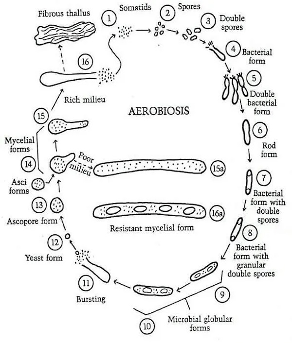
Apparently, Rivers was unfamiliar with the fact that this bacterial life cycle process, known as pleomorphism, was observed by many researchers such as Antoine Bechamp, Günther Enderlein, Royal Raymond Rife, and later by many others with the use of dark field microscopy. That bacteria are pleomorphic entities, i.e. having the ability to assume different forms, is an established fact.

VIRUSES AS FILTERABLE FORMS OF BACTERIA. For a long time a few investigators have held that certain virus diseases are induced by ordinary bacteria. Now that attention is being focused on filterable forms of bacteria, workers in increasing numbers (128, 131, 132, 134, 135) are adopting the belief that viruses are merely filterable, invisible, and noncultivable elements of ordinary bacteria. It has been claimed, and evidence of a kind has been offered to substantiate the assertions, that the bacteriophage (165, 166) is a form in the life cycle of lysogenic bacteria, that the viruses of yellow fever (131, 134, 135) and hog cholera (134, 135) are invisible forms of Leptospira icteroides and B. suipestifer respectively, that the etiological agent of scarlet fever (134, 135) is a filterable form of hemolytic streptococci, and that the incitants of poliomyelitis, epidemic encephalitis, fox encephalitis, common colds, measles, and influenza represent certain stages in the life cycle of green streptococci (131). Without going into details of the available knowledge of bacterial life cycles and their invisible and noncultivable forms, one can say that proof of many of the claims regarding them is lacking. In fact, if certain reports are correct, some of the filterable forms of bacteria are much smaller than are many of the viruses. Kendall (131) recently stated that “egg white, filtered through Berkefeld W filters (after dilution with sterile physiological saline solution) is rarely sterile.” Such a statement raises embarrassing questions for workers in the virus field because many viruses will not pass through W filters. Since the existence of bacterial life cycles is doubtful, it seems unwarrantable to offer the presumptive filterable forms of them as evidence upon another unsolved problem, the nature of the viruses.

The thing to notice in this next section on physical and chemical agents is, once again, the often contradictory nature of the evidence presented by different researchers. One researcher would find a certain chemical that had an effect on the “virus,” while another researcher would state otherwise. Some viewed that chemical tests proved “viruses” were protozoa. Others felt that their tests proved the “virus” was an enzyme. Sanderson showed that bacteriophages were not killed by successive freezing and thawings and believed that they were unliving. However, Rivers showed that bacteriophages can be killed by repeated freezing and thawing, thus contradicting Sanderson’s interpretation. Ultimately, Rivers concluded that, regardless of the number of tests with chemical and physical agents that had been devised as criteria for the presence of life or to define the nature of “viruses,” not a single one of them was found to be satisfactory.

EFFECT OF PHYSICAL AND CHEMICAL AGENTS ON VIRUSES. Many years ago it was discovered that bile and saponin are injurious to protozoa but with a few exceptions are innocuous for bacteria. Consequently, when the question of the nature of viruses began to attract attention, tests were made to determine what effect bile and saponin have on these incitants of disease. Many viruses, e.g., rabic virus (141, 144), were found to be inactivated and because of this fact certain workers concluded that they are protozoa. Sufficient exceptions, however, have been encountered to invalidate the test as a means either of separating bacteria from protozoa or of defining the nature of viruses. The agent causing Rous’ sarcoma (140) is more resistant to ultraviolet light than are bacteria, and Murphy (220) considers this fact as evidence in favor of his hypothesis of the enzyme-like nature of the virus. On the other hand, bacteriophage (139), the living nature of which many doubt, is just as sensitive to ultraviolet light as are bacteria. Sanderson (153), using a temperature of -78°C., found no diminution in the titer of two strains of bacteriophage subjected to 20 successive freezings and thawings. Since bacteria and cells are killed by repeated freezing and thawing, he concluded that bacteriophage must be something other than a living organism. Rivers (151) showed, however, that colon bacilli, Virus III, vaccine virus, herpetic virus, bacteriophage, complement, and trypsin are all either killed or inactivated by repeated freezing (-185°C.) and thawing and that, as might be expected, some of the agents are more resistant than are others. Hence it is obvious that destruction or inactivation of an active agent by repeated freezing and thawing is not evidence that it possesses life. The observations on heat, desiccation, oxidation, and the effect of dyes have likewise yielded no convincing evidence concerning the nature of viruses. Thus it appears that a number of tests with chemical and physical agents have been devised as criteria for the presence of life or to define the nature of viruses, but no one of them has been found satisfactory.

The spontaneous generation of “viruses” by the host is a concept that defeats the idea that these entities are exogenous outside invaders. If something like a bacteriophage can be produced by a normal bacterium without any external phage present, it shows that these entities arise from a process initiated from within the organism. Rivers noted that Hadley and his co-workers stated that it was possible to obtain bacteriophage from normal bacterial cultures by means of enforced dissociation. Thus, no external source of phage was necessary. Nobel Prize-winning immunologist Jules Bordet was able to do the same, as did other researchers. Rivers presented a few scenarios where “viral” diseases could be induced by injecting toxic substances such as tar and arsenic into chickens, as well as an instance where a tumor-producing extract could be obtained from healthy chickens. While Rivers thought that the interpretation of the evidence was potentially fundamental to biology, he excused it as being due to contamination by the researchers working in labs with similar materials as well as the possibility that “latent viruses” were hiding within the healthy hosts.

SPONTANEOUS GENERATION OF VIRUSES. The origin as well as the nature of viruses constitutes a question of interest. The intimate relation between these active agents and their host cells has induced more than one investigator to view the host cell as the source or origin of viruses. Indeed, reports of experimental work have appeared leading to claims that normal cells have been induced to manufacture certain viruses. According to Carrel (156, 157), minced chick embryo mixed with tar, indol, or arsenic and injected into normal chickens in a small percentage of instances gives rise to tumors resembling Rous’ sarcoma no. 1 and transmissible by cell-free filtrates. Fischer (163) by treating cultures of normal cells with arsenic obtained on one occasion a filterable agent capable of causing tumors. Carrel was unable to confirm Fischer’s work. Murphy (52, 167), by means of a method the details of which have not been described, reported that he was able to extract a filterable tumor-producing agent from the gonads of normal-appearing Plymouth Rock roosters. Recently, Hadley and his co-workers (166) stated that it is possible to obtain bacteriophage from normal bacterial cultures by means of enforced dissociation. Although no worker in this field has claimed to have generated living organisms from inanimate matter, it appears that a few believe that they have by certain manipulations induced cells to yield substances which possess some of the attributes of life, notably that of increasing without limit.

The observations described above are suggestive, and, if confirmed and found to warrant the interpretation given them by Carrel, Murphy, Fischer, and Hadley, will prove to be of fundamental biological importance. Unfortunately, however, all of the experiments yielding the observations were actively referred to were conducted in laboratories where workers engaged in the study of agents similar to those supposedly brought into existence. In such laboratories and with such materials it is always difficult for one to rule out the possibility of contaminating normal animals, tissues, bacteria, emulsions, and filtrates. This fact has long been appreciated by workers in vaccine virus laboratories and it delayed the acceptance of the experimental transformation of smallpox virus into vaccine virus. Therefore, experiments of the nature described should never be conducted in rooms used for the study of agents similar to those for which a search is being made. The workers who believe that they have induced viruses to come into existence have not excluded the possibility of the preexistence of latent viruses or of small amounts of virus in the supposedly normal embryos, gonads, chickens, and bacterial cultures utilized in the experiments. This possibility is emphasized by Flexner’s (164) work on poliomyelitis, for he was able to demonstrate the presence of virus in the nasal washings from normal contacts. The possibility outlined is further emphasized by Andrewes and Miller’s (155) experience with Virus III in rabbits, by Cole and Kuttner’s (158) work with the salivary-gland virus in guinea pigs, and by the work upon virus carriers in general among animals, plants (168) and bacteria.

CGI (Computer-Generated Imagery): The only way you will ever see “antibodies” attacking “viruses.”

Rivers next discussed “immunity” in relation to establishing the nature of “viruses.” It is important to note that, regarding antibodies and “immunity,” researchers are utilizing one hypothetical entity in order to define another. While Rivers spoke as if the antibody and antigen concepts are established facts, he remarked that if the concept of the nature of antigens is correct, “viruses” are proteins or are closely linked to proteins. Thus, the interpretation of the nature of the “virus” rests upon the correctness of the nature of the antigen concept. He felt that the rise of these (hypothetical) antibodies that differed between host cell and antigen adduced (led one to believe) the exogenous rather than the endogenous origin of the “viruses.” Regardless, Rivers admitted that the mode of action of neutralizing antibodies was not clearly understood, and when speaking of antibodies causing flocculation (clumping together), he shared that various researchers noted the “immunological” phenomena in “virus” maladies are comparable to those induced by toxins. While Rivers felt that “immunological” observations were important, he admitted that this method of approach had not brought about a definite solution to the problem of the nature of “viruses.”

IMMUNITY. Most virus diseases lead to a marked and lasting immunity in recovered hosts. Not only are the but in their sera antibodies capable of hosts refractory to reinfection neutralizing the viruses are demonstrable. What bearing have these facts upon the nature of viruses? In the first place, it is certain that viruses are highly antigenic. Furthermore, if our concept of the nature of antigens is correct, the viruses are proteins or are closely linked to proteins. Moreover, the agents are not only antigenic, but they give rise to antibodies different from those excited by proteins of the host cells. This is true even of the bacteriophage (188). These facts have been adduced as evidence of the exogenous rather than the endogenous origin of the viruses. Thus, the antigenic nature of viruses appears to be prejudicial to the idea that they are products of cellular activity. The notion, however, that a lifeless agent may be injurious to the cell creating it and that it may induce immunological responses independent of those excited by the cell, loses some of its fantastic qualities when one considers the well-known facts that lens protein is not species specific but organ specific and that sympathetic uveitis in the uninjured eye is caused not by microorganisms but by the reaction of the body to substances derived from injured cells of the other uveal tract.

In addition to the neutralizing antibodies, whose mode of action is not clearly understood, complement-fixing antibodies and antibodies causing flocculation in virus emulsions have been described. Schultz and his associates (191-195) contend that the latter types of antibodies are not excited by viruses and that the immunological phenomena in virus maladies are comparable to those induced by toxins. In spite of their contentions, sufficient evidence has been adduced by different workers to make it more than likely that certain virus diseases lead to the production (176, 177, 180, 199) of the antibodies mentioned. Furthermore, Ledingham (184) has recently demonstrated that Borrel bodies in fowl-pox and Paschen bodies in vaccinia are specifically agglutinated by antifowl-pox and antivaccinal sera respectively. The results of these experiments indicate to Ledingham that the elementary bodies are living organisms and represent the virus. There is no reason to doubt that specific agglutinations of the bodies occurred in the manner described by Ledingham, and one cannot deny that such a phenomenon is presumptive evidence of the organismal nature of the bodies. Yet one dare not say categorically that his experiments are unequivocal evidence that the elementary bodies represent virus alone, because it has been shown by Jones (182, 183) that collodion particles treated with a variety of proteins and then thoroughly washed are specifically agglutinated by the proper antisera. Thus, the Borrel and Paschen bodies without being organisms yet having virus adsorbed on them might nevertheless be specifically agglutinated by appropriate antiviral sera.

Gye (181) states that Rous virus repeatedly injected into alien hosts excites two groups of antibodies, one of which acts on the virus itself, while the other operates on the “specific factor” derived from the host cell. According to him, either set of antibodies inactivates the virus. This fact is offered by him as further evidence of the dual nature of the causative agent of fowl tumors. Murphy (189) and Sittenfield (196-198) have reported the presence in Rous sarcoma of a substance that inhibits the action of the etiological agent, and the first mentioned worker is of the opinion that the “inhibitor” differs from ordinary virus antibodies. The presence of this “inhibitor” together with other phenomena has induced Murphy (220) to believe that immunity to the Rous agent is unlike that observed in virus maladies and lends evidence to his view that the Rous agent is not a virus. Inhibiting substances, however, have been obtained from tissues infected with viruses, for example, a substance restraining the action of rabic virus has been demonstrated by Marie (186) in the brains of rabid animals. Furthermore, Andrewes’ (172, 173) work appears to indicate that the immune responses excited by the filterable agents of fowl tumors may not be unique and may possess much in common with those encountered in other virus diseases.

From what has been said, it is obvious that immunological phenomena are playing an important role in discussions concerning the nature of viruses. As yet this method of approach has not brought us to a definite solution of the problem.

Rivers finished up his review on the nature of “viruses” by presenting the various differing interpretations on the concept of the “virus.” These invisible entities were regarded as either:

  1. Living contagious fluids
  2. Oxidizing enzymes
  3. Protozoan parasites
  4. Inanimate chemical substances
  5. Minute living organisms (related to bacteria)

Rivers noted that depending on the researchers, the rabies “virus” was either an enzyme, a parasite, a protozoon, or an unknown living organism. He stated that researchers were divided over whether bacteriophages were an inanimate agent or a living organism. The fowl-pox “virus” was thought of as either a protozoan parasite, a nucleoprotein poison manufactured by “infected” cells, or a minute coccoid organism capable of regeneration in parasitized cells. The agent associated with Rous sarcoma was either animate, a living organism mixed with an inanimate substance, an enzyme-like substance, or a transmissible mutagen.

Rivers highlighted these numerous competing concepts in order to show how radically different the ideas concerning the nature of “viruses” are from one another. He then proceeded to explain the main conceptions of “viruses,” with the first two scenarios explaining how a stimulus induces a normal cell to create a substance X, which may either remain free or become closely bound to a part of the cell. In Rivers’ third example, which he considered the most popular, X is a minute living organism that enters cells, multiplies, and produces disease. Thus, there is a distinct difference where X is considered an inanimate substance that results from cellular perversion in the first two scenarios, while X is viewed as an autonomous organism in the last scenario. Regardless of the scenarios that Rivers provided attempting to explain “viral” formation, he admitted that there was no unequivocal evidence of the validity of any of these concepts.

CONCEPTS OF THE NATURE OF VIRUSES. A review of the data by means of which one arrives at a concept of the nature of viruses has been presented. Now it will be interesting to see what notions certain workers have concerning some of them.

Beijerinck (202) considers the virus of mosaic disease to be a living contagious fluid; Woods (228), an oxidizing enzyme; Goldstein (212), a protozoan parasite; Vinson (67), an inanimate chemical substance. Most workers, however, believe that it is a minute living organism.

Hijgyes (216) is of the opinion that the incitant of rabies is an enzyme or “alternatively, that the tissues themselves might spontaneously become virulent as the result of changes in their chemical composition.” At one time Remlinger said, “The rabies virus, which is at once filterable, diffusible and capable of reproducing the disease from case to case, appears to occupy a place midway between the microbes and the diastases.” Recently, however, he (221) has published an article on the evolution of the parasite of rabies. Levaditi (118) and others (121, 225) have presented evidence in favor of the idea that the causal agent is a protozoon. The majority of investigators hold the concept that the incitant is a living organism whose nature is not definitely known.

Numerous workers believe that the bacteriophage is an inanimate agent, while others are convinced that it is a living organism. Ideas, however, concerning the nature of the inanimate transmissible substance or the animate organism vary. For details of the different concepts one is referred to papers by Twort (226, 227), d’Herelle (103), Bordet (203), Bronfenbrenner (95), Burnet (206), and Hadley (165, 166).

The incitant of fowl-pox has been described by certain investigators as a protozoan parasite. Sanfelice (222, 223) suggested that it is a nucleoproteid poison manufactured by infected cells. Borrel (log), Goodpasture (124, 125), and Ledingham (184) hold that it is a minute coccoid organism capable of regeneration in parasitized cells.

Rous and others are prepared to entertain the idea that the causal agent of Chicken Tumor No. I is animate. Gye (215) believes that it consists of two factors, one of which is a living exogenous organism, the other an inanimate specific factor derived from infected cells. Murphy (52, 55), at one time, spoke of the Rous agent as an enzyme-like substance. Recently, however, he (220) has compared it to filterable substances capable of transforming melitensis (204,205) into paramelitensis organisms and of converting one type specific pneumococcus (201) into another type specific form. In regard to the matter he says (22O), “Thus we have a group of agents, products of specialized cells capable of conferring the peculiar type quality to undifferentiated cells of the same species which, in turn, may produce the active factor and transmit this to their descendants.” For this type of agent he proposes the name transmissible mutagens.

Sufficient ideas concerning the nature of viruses have been cited to illustrate how radically some differ from others. Many of them, particularly the ones dealing with the origin and reproduction of inanimate substances that behave in a manner similar to that of living organisms, lack precision. In a general way, however, the different concepts can be arranged in groups and it seems advisable to state and to portray diagrammatically several of the popular ones.

According to one conception, certain stimuli produce changes within cells that are inherited by daughter cells. Once the mutations occur, cells of the new type continue to be formed though the stimuli disappear. No agents separable from the cells are demonstrable, and immunological phenomena in this type of disease differ from those observed in virus maladies. Ordinarily this idea of the causation of disease and the concepts concerning the nature of filterable viruses are not grouped together. Yet in some respects they are not dissimilar and many hold the view that malignant neoplasms arise in some such way. See figure 1.

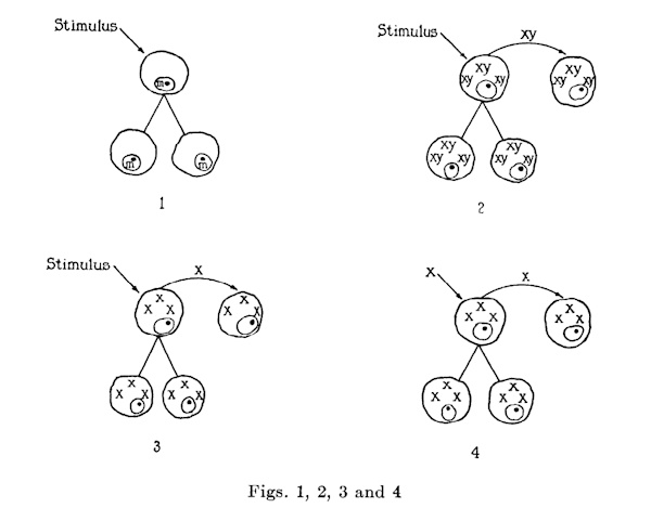
Another notion is that appropriate stimuli induce normal cells to make a substance x which is closely bound to parts y of the cells. Thus an xy complex is formed. This complex, separable from the cells, yet capable of inciting its own production by them, either passes directly into daughter cells, or, having become extracellular, enters another set of normal cells. The xy complex is antigenic, and cells freed from it presumably become normal again. See figure 2.

Still another idea is that certain stimuli incite normal cells to produce a substance x which is not closely bound to parts of the cells, X, separable from cells, yet capable of impelling its formation by them, either passes directly into daughter cells, or, having become extracellular, enters a new group of normal cells. X is antigenic and cells freed from it presumably become normal again. See figure 3.

Finally there is the concept most generally held that x is not a product of the perverted activity of cells but is a minute living organism. X enters cells, multiplies, produces disease, is separable from cells: and is antigenic. Cells freed from it presumably become normal again. At times, x is absorbed by particles y of host cells and evidences of an xy complex are obtained. See figure 4.

For practical purposes it makes little difference which one of the last three concepts is accepted. Theoretically, however, x of the second and third conceptions is quite different from x of the fourth. In the second and third, x, a product of cellular perversion, is an inanimate agent, while in the fourth it is an autonomous organism. No unequivocal evidence of the validity of any of the concepts has been adduced.

Rivers concluded by acknowledging the confused state of the evidence concerning “viruses,” noting that this confusion had made it exceedingly difficult to define their nature. He felt that the easiest way out of their dilemma would be to accept “viruses” as minute organisms. However, Rivers warned of quickly accepting presumptive evidence as “viruses” may be either minute organisms, forms of life unfamiliar to us, inanimate transmissible incitants of disease, or all of the above.

Conclusion

The confused state of our knowledge of the viruses at the present time makes it exceedingly difficult to define the nature of these active agents. The easiest way out of the dilemma, however, would be the acceptance of the presumptive evidence that viruses are minute organisms. Yet the easiest way and the one that best fits the experiences of the day may not be the right one. Furthermore, excessive skepticism and the habit of too readily accepting presumptive evidence are equally productive of sterility. Unless viruses represent a form of life unknown to us, proof of their living nature would not be a striking discovery. If, however, some of them are not animate, absolute proof of such a fact would be of fundamental biological importance. Therefore, care should be exercised that immoderate skepticism on the one hand, and the mental satisfaction secured by accepting presumptive evidence on the other, do not dull our efforts to obtain a better understanding of the viruses, some of which may be minute organisms, while others may represent forms of life unfamiliar to us, while still others may be inanimate transmissible incitants of disease. In any event, we are face to face with the “infinitely small in biology,” and, if there be a sharp demarcation between life and death, then scientists, investigating the nature of viruses, are working near the line that separates infinitely small living organisms from inanimate active agents.

https://www.nature.com/articles/145853d0

From these two presented articles from two different points in time in the history of virology (Rivers in 1932 and Summers in 2014), it should be clear why it is difficult for virologists to define the nature of the “virus.” Researchers needed to invent, and then continually reinvent, the nature of the “virus” as the foundation that virology is built upon is conceptually weak. It is full of contradictions that have cracked the very infrastructure that was put in place. There were never any submicroscopic entities that were being studied by the various researchers over the last century. As there were no “viruses” to study and characterize, there was no agreement at all amongst the various researchers as to the nature of the invisible concept crafted inside of their minds. They had tricked themselves, through shoddy indirect pseudoscientific evidence, into believing that they were studying something real based upon lab-created effects without an identifiable cause. This is why the “virus” has been continually defined for what it isn’t, rather than for what it supposedly is. The magical “virus” skirts the line between life and death, microbe and molecule, enzyme and ferment. It is unlike anything else seen in nature, and for this very reason, its nature remains mysterious and incomplete. This should be the very first clue that there is nothing scientific about the “virus,” as science only deals with the natural world and its phenomena, not the supernatural. However, within the supernatural realm is where the “virus” concept will remain, ready and waiting to be reinvented upon the arrival of the latest technology for the next best indirect measurement. This will be utilized to continue fooling the researchers, as well as the public that blindly trusts in them to know better, that these fictional entities exist in nature, when, as Thomas Rivers kindly pointed out, “viruses” have never once been observed there. Thus, the nature of the “virus” will continue to remain merely an invention of the imagination of the most ardent admirers of these invisible boogeymen—the virologists.

This article originally appeared on ViroLIEgy’s Antiviral Substack.


Connect with Mike Stone website | substack

Cover image credit: flutie8211
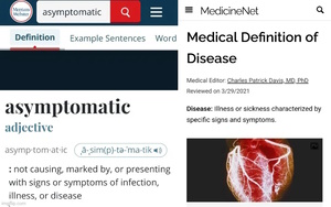
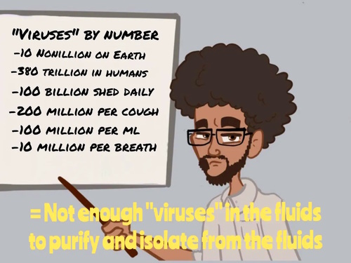
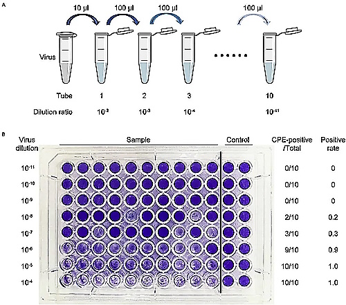
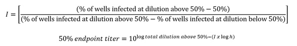
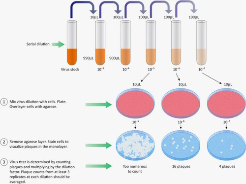
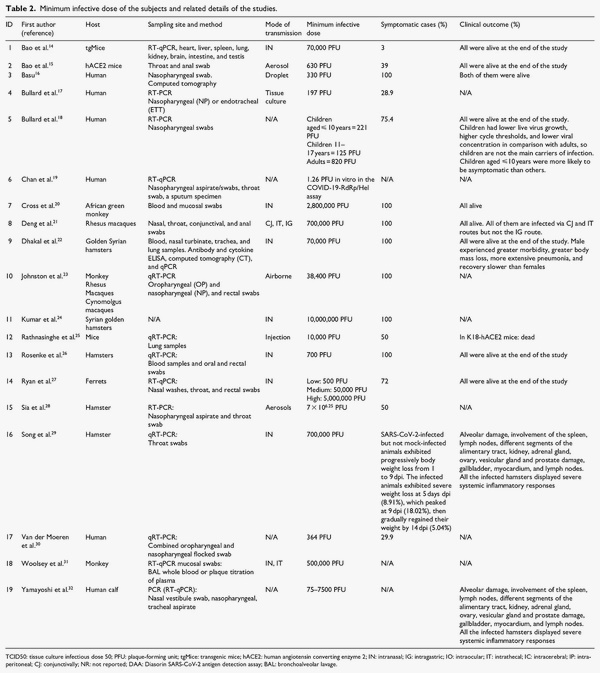
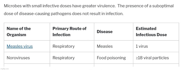
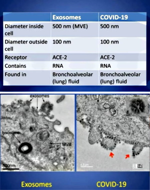
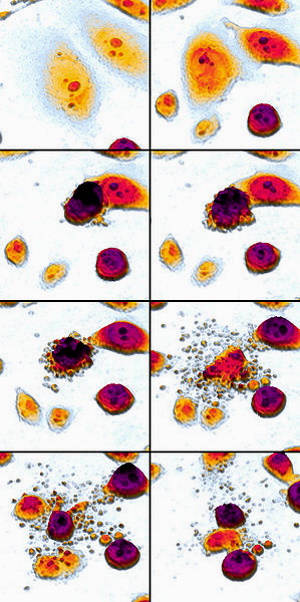
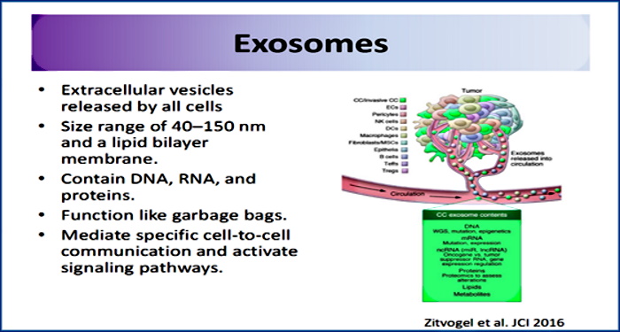
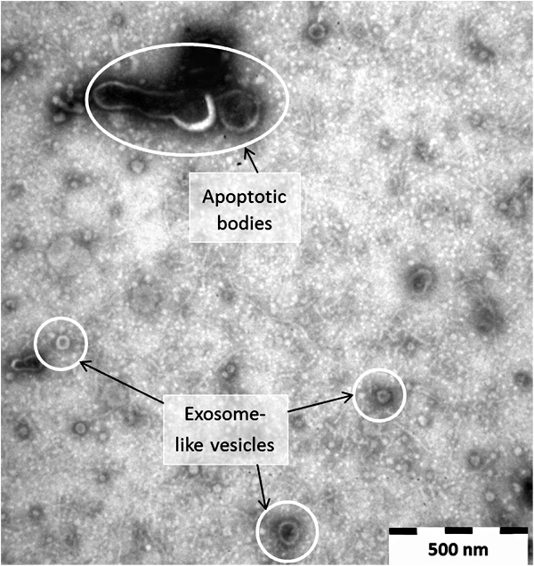
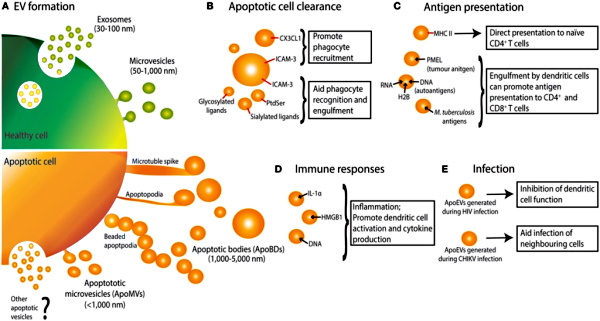
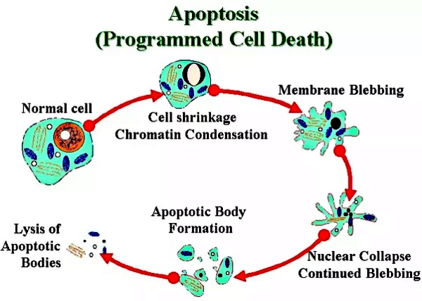
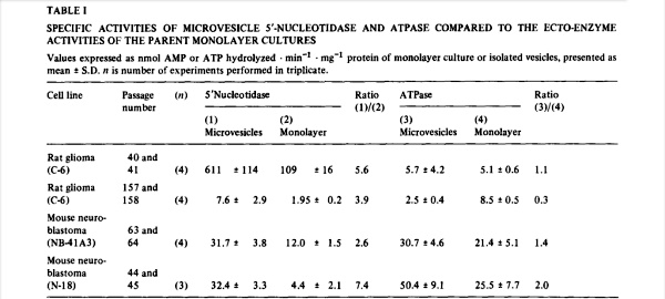
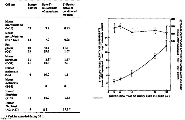
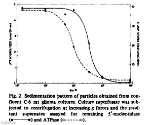
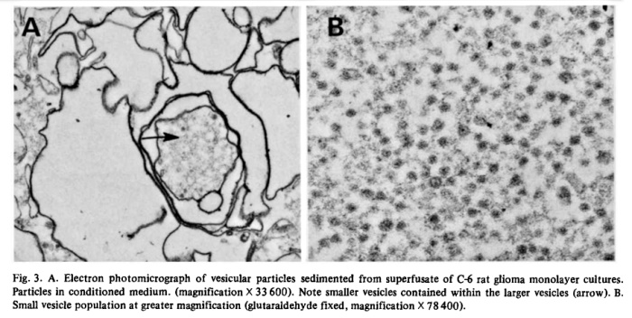
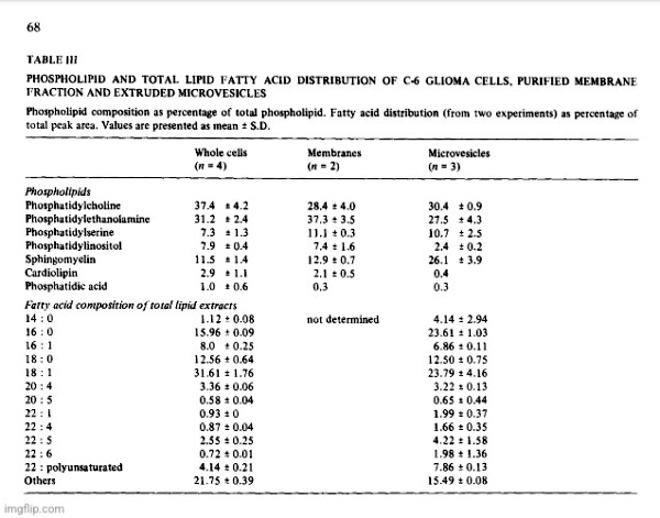
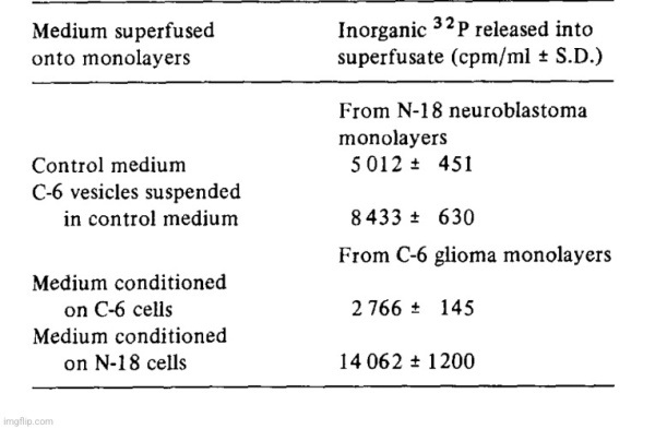
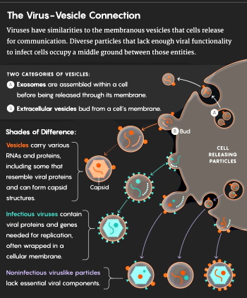
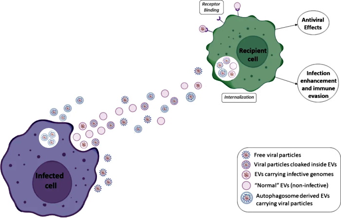
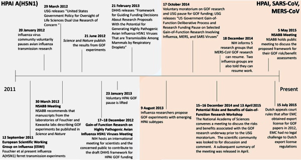
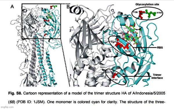
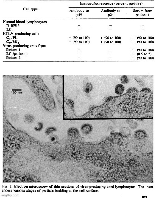
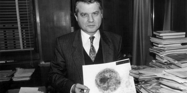
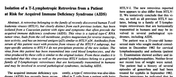
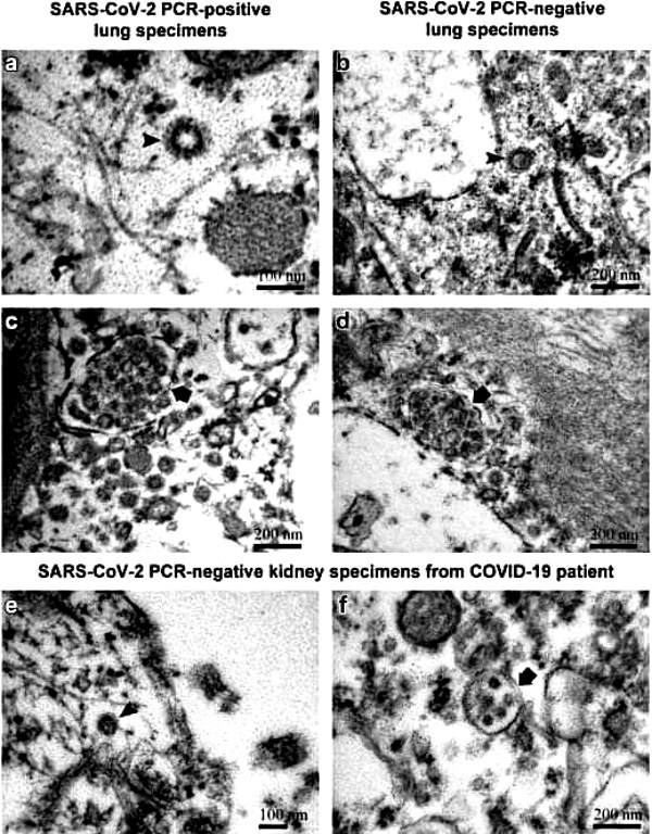
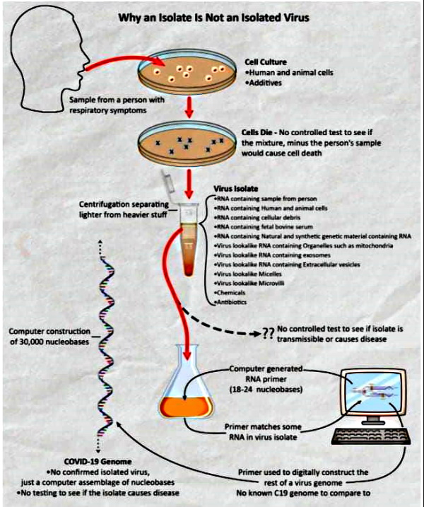


The Healthy Sick: Mike Stone on the Pseudoscience Behind the Idea of Asymptomatic Carriers of Disease

The Healthy Sick: Mike Stone on the Pseudoscience Behind the Idea of Asymptomatic Carriers of Disease


“…This is a pseudoscientific concept that is also an oxymoron as two entirely contradictory terms were put together in order to create this illogical state. Asymptomatic is characterized by a lack of signs and symptoms of illness, whereas disease is characterized by signs and symptoms of illness…

Asymptomatic carriers are nothing but healthy people who have been labelled with disease minus signs of any disease who are then told that they can infect others. They are treated as a sick individual based upon results generated using fraudulent tests.

[…]

The “viral” theory is a load of BS, and there is no such thing as a healthy sick person capable of transmitting disease. We have no reason to fear the walking healthy.”

The Healthy Sick
Fear the walking healthy. 

by Mike Stone, ViroLIEgy
May 5, 2023


“In areas where there are limited number of new cases, State or local public health officials may request to test a small number of asymptomatic ‘healthy people,’ particularly from vulnerable populations”

-CDC Revised Guidelines August 2020 (source)

In the not so distant past, when we walked around feeling healthy without any symptoms of disease, most of us would consider that we were, in fact, free of any disease. There would be no thoughts about going to the doctor for a PCR test in order to determine whether or not we were unknowingly a walking talking “virus” spewing host harboring billions of “infectious” particles capable of transmitting disease to our loved ones. We would not subject ourselves to quarantines and daily testing due to the remote possibility of being around someone who tested positive with symptoms, let alone for anyone testing positive without any signs of disease. We did not go around covering our faces with masks out of fear that those around us may be silent spreaders. We didn’t bust out our rulers in order to measure 6 feet of distance between us and another living soul. None of these irrational actions were ever even a glimmer of a thought until the well-orchestrated fear propaganda campaign promoted the pseudoscientific concept of the asymptomatic carrier of disease and catapulted it into the public consciousness.

Even though this idea has been effectively weaponized against us over the last few years, it is not a new one. In fact, as will be shown later, the notion of the asymptomatic carrier began at the same time germ theory was born. The idea is that one can be silently harboring and able to transmit a pathogen without displaying any symptoms of disease whatsoever. This has resulted in the highly illogical creation of asymptomatic disease:

What Does it Mean to Have an Asymptomatic Disease?

“Asymptomatic disease is where a person is infected with a disease (or develops a disease; diagnosed) but fails to display any noticeable symptoms.”

Asymptomatic until symptomatic – silent diseases

“Many diseases and infections can be asymptomatic, including those that may be potentially fatal in some people. These include (but are not limited to): tuberculosis, breast cancer, endometriosis, HIV/AIDS, herpes, hepatitis, chlamydia, hypertension, common colds/flu, and type-2 diabetes mellitus. Many of these conditions remain largely asymptomatic until very advanced disease stages when they suddenly become symptomatic. Others can remain more or less asymptomatic throughout their disease course.”

“Infectious diseases can also be completely asymptomatic (with no symptoms ever manifesting), particularly in younger and healthier individuals. For example, hepatitis (hepatitis C) infections can take up to 6 months to develop, and even then, approximately 80% of infected individuals may not experience any symptoms. Other examples include cholera, herpes, measles, and rubella which can be completely asymptomatic.”

“In summary, asymptomatic disease refers to diseases and infections which do not lead to any symptoms in patients (subclinical) for the whole disease course or until they develop symptoms in which the asymptomatic phase is referred to as pre-symptomatic.

In many respiratory infections including COVID-19, asymptomatic disease is common and may be a source of transmission within the community, though more research is needed to establish the exact contribution asymptomatic transmission has on the community rates of infection.”

(source)

As can be seen, many so-called “infectious diseases” are said to be asymptomatic. If one is labelled as asymptomatic, one never develops the disease at any point in time even though they are diagnosed with asymptomatic disease. This is a pseudoscientific concept that is also an oxymoron as two entirely contradictory terms were put together in order to create this illogical state. Asymptomatic is characterized by a lack of signs and symptoms of illness, whereas disease is characterized by signs and symptoms of illness. One can not have disease if one is not displaying signs of disease:

Asymptomatic carriers are nothing but healthy people who have been labelled with disease minus signs of any disease who are then told that they can infect others. They are treated as a sick individual based upon results generated using fraudulent tests. In the past, most would have scoffed at this idea and never willingly subjected themselves to quarantines and further testing. In fact, they would have never tested to begin with. However, in the face of a “pandemic” with a “novel virus,” many lined up for the mass testing agenda in order to ensure that they were amongst the “uninfected.” This willingness to subject to testing despite a clear lack of symptoms was primarily driven by fear. This old concept was thrust onto a frightened population and then ramped up in a way that had never been done so before.

In order to understand why there was never any reason to ever participate in this irrational belief of such a ridiculous concept, let’s examine how the asymptomatic carrier first came about at the dawn of germ theory. We will then examine how this idea was weaponized against the public during the “pandemic” despite a complete lack of any scientific evidence in support of the asymptomatic disease carrier.

When German bacteriologist Robert Koch was looking for the causative agents of certain diseases in the late 1800’s, he formulated a series of four logical requirements that needed to be met in order for anyone to claim that a certain microbe caused a specific disease. These were as follows:

  1. The microorganism must be found in abundance in all hosts suffering from the disease but should not be found in healthy hosts.
  2. The microorganism must be isolated from a diseased host and grown in pure culture.
  3. The cultured microorganism should cause the same symptoms of disease when introduced into a healthy host.
  4. The microorganism must be re-isolated from the inoculated, diseased experimental host and shown to be identical to the original causative agent.

While these logic-based postulates were accepted by and large within the scientific community, Koch quickly discovered a problem with his very first criterion. Whether it was tuberculosis, typhoid, malaria, or cholera, the microbe that he was claiming as causative agents were regularly found in healthy individuals. Thus, Koch was unable to satisfy his very own first Postulate. However, rather than realize that his criteria had worked as he had envisioned and had actually ruled out bacteria and other microbes as a causative agent of disease, Koch allowed for himself and others to bend not only his first postulate, but the others as well. Allowing for the bacteria and other microbes claimed to be causative agents of disease to be found in those without disease lead to the creation of the illogical concept that became known as the asymptomatic carrier of disease. Koch’s entire claim to fame rested entirely on the perception that he was a microbe-hunter. Bending his own rules saved Koch from giving up his prestige, kept his findings intact, and helped to establish the germ theory on unfalsifiable pseudoscientific grounds.

Koch’s idea of asymptomatic “infection” received a big push shortly afterwards when the media released propaganda promoting the idea of an asymptomatic carrier in 1907 by targeting an Irish immigrant by the name of Mary Mollen. Mary was a cook for wealthy families and ended up employed by banker Charles Henry Warren when he rented a summer home for himself and his family. When 6 of the 11 family members came down with the symptoms of typhoid fever over the last week of August, the property owners feared that no one would rent the house again if they believed that the property was the source of the outbreak. A man named George Roper was hired to investigate the situation and he came to the conclusion that it was Mary who had passed on the bacteria to the family through her cooking. This led to a modern day witch-hunt for Mary who refused to believe that she was the source of illness. Sadly, Mary was eventually involuntarily quarantined for the majority of the rest of her life. This ordeal led to Mary being notoriously and unfairly known by the moniker Typhoid Mary, even though many of her stool samples came back negative for the bacterium:

Typhoid Mary: the Tragedy of Mary Mallon

“On 11 November 1938, a 69 year old Irishwoman died on North Brother Island, New York. She had been held in isolation for 23 years, yet she had not been charged or convicted with any criminal offence.

Mary Mallon was born in Cookstown, Ireland in 1869. She immigrated to America when she was a teenager and found employment in domestic service. She developed an aptitude for cooking, and as this paid more than basic service, Mary accepted several jobs as a cook for the wealthy. In 1906, Charles Henry Warren, a New York banker, rented a summer home for himself and his family on Long Island. Mary Mallon was engaged as a cook for the duration of their stay. From the end of August, one by one people began to fall ill with typhoid fever, in all, six of the eleven occupants of the house developed typhoid fever.

The owners of the property feared that they would be unable to secure further tenants if the public believed that the source of the outbreak was their property and so hired Dr George Soper to investigate the cause. Soper came to the conclusion that Mary Mallon was to blame for the spread of disease. Hindering his efforts, Mary had left their employment three weeks after the outbreak. Soper started to investigate the previous situations held by Mary Mallon. From 1900 to 1907 there had been seven jobs where, it was reported, somewhere between twenty-three and thirty-eight people became ill and one person, a child, died. Soper believed that Mary was the source of typhoid fever that had followed her employment history, but he needed biological samples to affirm his hypothesis.”

“The Greater New York Charter allowed for ‘all reasonable means for ascertaining the existence and cause of disease’. It essentially gave health officials the authority to remove Mary Mallon and quarantine her against her will. After two years of isolation, with only a dog for company, Mary sued the health department. They had tested her stools approximately weekly and 120 out of 163 samples proved positive. Yet Mary countered with her own private analysis, sampled over the preceding year, all coming back negative. Mary’s laboratory results proved for her, her healthy status and she failed to understand that she was diagnosed a healthy typhoid carrier. She was arguably the first person identified as such, and having not been charged with a criminal offence she felt it was barbaric to be treated like a criminal (and a ‘leper’) when she was innocent of any crime.”

(source)

Mary was falsely quarantined against her will due to one man’s suspicion and hypothesis that rested solely on correlation equaling causation. No scientific experiments were ever carried out proving that Mary was spreading disease to her patrons. As with all claims of asymptomatic transmission, it was a circumstantial case built upon faulty epidemiological data. George Roper is the man who ultimately condemned Mary by labeling her as the cause without any scientific evidence proving his hypothesis. Based upon his own words presented below, he assumed certain premises, such as the bacterium should be in the urine (which it was not) and in the feces. He claimed that stool examinations only failed twice over the course of two weeks to find the bacterium. However, he later recounted several instances of failure to detect the bacterium over the course of several months. In the summer months, few bacterial colonies were found and in the month of July, there were five consecutive negative tests. During the month of August, no typhoid was ever found in Mary’s stools. In September, they began to appear again. However, from September 11 to October 14, 1907, the stools failed to yield any typhoid bacilli. From October 16, 1907, to February 5, 1908, weekly examinations of the stools showed anywhere from 25 to 50 percent “typhoid-like” colonies on the culture plates. There were two instances within that period where no bacilli were found. Taking into account that Mary’s own independent lab results showed that no bacilli were found within her stools, Soper’s consistently contradictory evidence should have been questioned.

After recounting these failures, Soper shared his thoughts on how Mary transmitted the bacterium through having not washed her hands properly while preparing the food. He based his conclusion upon his interviews where he stated that no housekeeper ever told him that Mary was a clean cook. He did not say whether he asked them or not or rather just assumed that their lack of addressing it was proof that Mary was unsanitary. Soper then stated that, in the most thorough “investigation,” he believed that the bacterium was carried from Mary’s hands to the people who ate ice cream containing cut-up peaches that she had prepared.  Again, no evidence was provided beyond his belief. Soper was amazed that no one had ever discovered an asymptomatic carrier in America before him. Interestingly, Soper revealed that he was long interested in the transmission of typhoid fever and knew of Robert Koch’s work. He stated that his interest in this area was longstanding and that Koch’s work was the basis for his own investigation. He admitted that he had read several papers on the probable role of healthy carriers in producing typhoid. Soper was made aware by Dr. Simon Flexner, of the infamous Flexner report, to some of these references after he had concluded his work on the Mary Mallon case. It is very clear that Soper went looking for evidence to fit his preconceived conclusions as to what the cause was. He was also potentially guided along the way:

The Military Surgeon Vol. XLV July, 1919 Number 1 Original Articles Typhoid Mary
By Major GEORGE A. SOPER

“It was expected by me that the germs might be found in the urine, but more probably in the stools. None was found in the urine. The stools contained the germs in great numbers. Daily examinations made for over two weeks failed only twice to reveal the presence of the Bacillus typhoid and on these occasions the sample taken was perhaps too small to reveal them. The blood gave a positive Widal reaction. The cook appeared to be in perfect health.

The feces were examined on an average of three times a week from March 20 to November 16, 1907, and in only a comparatively few instances did the investigators fail to find the bacilli. During the summer months the culture plates contained only a few typhoid-like colonies. In July there were five consecutive negative tests followed by a positive one.

During August the stool showed no typhoid; in September they began to appear again; from September 11 to October 14, 1907, the feces failed to yield typhoid bacilli. During this time the patient’s diet was carefully regulated and she was receiving mild laxatives.  On October 16, 1907, a very thorough test showed that the germs were again present. From October 16, 1907, to February 5, 1908, weekly examinations of the stools gave, with only two exceptions, from 25 to 50 per cent typhoid-like colonies on the culture plates. These exceptions were on November 13 and December 4, when no typhoid was found. The implication was plain. The cook was virtually a living culture tube in which the germs of typhoid multiplied and from which they escaped in the movements from her bowels. When at toilet her hands became soiled, perhaps unconsciously and invisibly so. When she pre-pared a meal, the germs were washed and rubbed from her fingers into the food. No housekeeper ever gave me to understand that Mary was a particularly clean cook. In the Oyster Bay outbreak, which was studied with more particularity than the others, the infectious matter is believed to have been carried from the cook’s hands to the people who were later taken sick by means of ice cream containing cut-up peaches.  Mary prepared this herself. In this instance no heat sterilized the washings from her hands. Mary Mallon was kept virtually a prisoner by the Department of Health for three years. At first she was held at the hospital for contagious diseases at the foot of East 16th Street, Manhattan; later she was removed to Riverside Hospital on North Brother’s Island in the East River, between Hell Gate and Long Island Sound.”

“The case is least remarkable for the reason that it was the first of its kind to be worked out in America. It is surprising that nobody bad discovered a carrier before. They are now known to be rather common.

Somewhat similar investigations bad been made in Germany) and I make no claim of originality or for any other credit in her discovery. My interest and experience in the epidemiology of typhoid had been of long standing. I had read the address which Koch had delivered before the Kaiser Wilhelm’s Akademie, November 28, 1902, and his investigation into the prevalence of typhoid at Trier 3 and thought it was one of the most illuminating of documents. In fact it had been the basis of much of tile epidemic work with which I had been connected.

Koch’s address was not the only one printed about this time to show that healthy carriers might exist and give rise to typhoid.  Conradi and Drigalski4 had anticipated Koch and it was probably on the suggestion contained in their paper to the effect that with their new culture medium they had found typhoid bacilli in the stools of several well persons that Koch’s flying laboratory was sent to Trier and the ground prepared for his Kaiser Wilhelm’s Akademic address.

In the Festschrift Zum SeclizigstenGeburstag von Robert Koch, which appeared in 1903, there are several papers on the probable role of healthy carriers in producing typhoid. About this time Kayser, Klinger and others were publishing in Arbeiten aus dem Kaiserlichen Gesundheit-smate reports of cases which they found to be due to persons whose condition was much like Typhoid Mary’s.  Dr. Simon Flexner kindly called my attention to some of these references after I had concluded my work on the Mary Mallon case.”

(source)

After the highly publicized Typhoid Mary case, this idea of asymptomatic carriers simmered in the background over the next century. While there were claims of such a state in certain diseases, this has never been scientifically proven. However, that did not stop Koch’s escape clause from taking a prominent role in the “Covid crisis,” primarily due to a mass testing campaign that was bound to identify positive cases in healthy people using fraudulent tests never calibrated and validated to purified and isolated “virus.” Although all PCR results are false-positives, we can see that even the CDC noted that testing people without symptoms generates false-positive cases. They stated as much under their PCR guidelines for pertussis when recommending not to test those without symptoms:

Diagnosis PCR Best Practices

“However, only patients with signs and symptoms consistent with pertussis should be tested by PCR to confirm the diagnosis. Testing asymptomatic persons should be avoided as it increases the likelihood of obtaining falsely-positive results. Asymptomatic close contacts of confirmed cases should not be tested and testing of contacts should not be used for post-exposure prophylaxis decisions.”

(source)

Thus, we can see that the CDC were well aware that testing people without symptoms will lead to an influx of cases labelled as asymptomatic “infections” when they are, in fact, not “infected” or diseased at all. This massive amount of asymptomatic cases of “SARS-COV-2” based upon fraudulent test results has cemented the illogical concept of the asymptomatic carrier into the minds of the populace. A timely December 2020 review, while reiterating the history of the asymptomatic carrier described above, pointed out the fact that even though asymptomatic infection and transmission has always been a concept waiting it the wings, it has only recently been thrust into the limelight with this “pandemic:”

Invisible epidemics: ethics and asymptomatic infection
History

“Dr Robert Koch was one of the founders of modern microbiology, and his work is particularly well known for a set of postulates (first published in 1890) linking microbes with the causation of infectious disease (Gradmann 2010). Though variously expressed, one of Koch’s initial postulates was that the microbe putatively responsible for a disease should be found in all people suffering from the disease, but not in healthy individuals (Gradmann 2010). Koch soon realised that this did not hold true in all cases, since many potentially pathogenic organisms are frequently found in healthy people. For example, Koch observed that asymptomatic carriers of cholera, typhoid, and malaria could spread these diseases to others, and he is credited for inventing the concept of the carrier state (i.e., in which healthy people asymptomatically carry an infection) (Gradmann 2010).

Public awareness of asymptomatic carriage of infection increased, especially in English-speaking countries, with media reporting of the case of Mary Mallon (known as “Typhoid Mary”) beginning in 1907. Mallon was a cook working in New York who, although showing no signs of typhoid disease herself, spread typhoid bacteria to many other people, resulting in several deaths (Brooks 1996; Soper 1939). For the general population, this revealed an important truth: that “persons, rather than things” (Soper 1939) were the source of many infectious diseases. Despite this Copernican revolution in public health (an epidemiological parallel of the microbiological revolution of germ theory), Mary Mallon and many others found it difficult to believe that healthy people could spread disease. Mallon repeatedly resisted public health restrictions and refused to believe she was infected or posed risks to others. She spent the latter years of her life living in public health confinement on North Brother Island, working as an assistant in the local infectious disease laboratory (Soper 1939).”

Implications for outbreaks, epidemics, and pandemics

“Asymptomatic infection was recognised to be a significant factor in the 2015–2016 Zika virus epidemic, particularly because many of those who were infected—including some women who acquired infection during pregnancy and gave birth to children severely affected by congenital Zika syndrome—showed few or no symptoms (Jamrozik and Selgelid 2018). Although less well recognised, transmission of asymptomatic Middle Eastern Respiratory Syndrome (MERS) coronavirus infection (perhaps both camel-human and human–human transmission) may play an important role in the epidemiology of MERS—which is all the more remarkable because people who develop symptomatic MERS infection have a high fatality risk of around 35% (Grant et al. 2019). Asymptomatic infection has also been reported for viruses closely related to the coronavirus that caused the earlier severe acute respiratory syndrome (SARS) epidemic. In one study from 2003, around 40% of Chinese wild animal traders had serological evidence of having been exposed to coronaviruses that closely resembled SARS-coronavirus, raising questions about whether people in high risk occupations should be screened for asymptomatic infection to detect potential “spillover” events of pathogens with epidemic potential (Guan et al. 2003). We initiated the November 2018 Brocher Foundation workshop upon which this Special Issue is based partly in light of the growing awareness of such cases of asymptomatic infection—and their ethical implications for policy and practice.

Since that time general awareness of asymptomatic infection has skyrocketed in light of its role in the coronavirus disease 2019 (Covid19) pandemic, in virtue of which the term ‘asymptomatic infection’ has become highly familiar to ordinary members of the general public. Early data, which were later widely confirmed, suggested that asymptomatic transmission of Covid19 occurs both in cases where the individual transmitting the virus goes on to develop symptoms later (i.e., they were “pre-symptomatic” at the time of transmission) and in cases where they never develop symptoms (Hu et al. 2020). Asymptomatic individuals can, under certain conditions, transmit to large numbers of other people (e.g., one person was shown to infect 71 others) (Liu et al. 2020). The overall degree to which asymptomatic transmission contributes to local Covid19 epidemics likely varies in different contexts and has not always been well-characterised (in part because of the difficulties of identifying all asymptomatic infections during an epidemic). In any case, asymptomatic transmission of Covid19 raises a number of ethical issues similar to those discussed above, including those related to the justification of public health interventions such as screening and isolation for asymptomatic cases.”

(source)

While the asymptomatic carrier was made a star of the “Covid” show in order to generate fear and drive compliance towards quarantines, lockdowns, social-distancing, and masking, the message has been entirely inconsistent throughout, and the lack of any valid scientific evidence proving such a carrier state was on full display from the very beginning. At a White House press briefing on January 28th 2020, the idea of asymptomatically transmitting the “novel coronavirus” was floated out there as a possibility. The CDC claimed to have heard reports about asymptomatic cases but had not seen any of the data. At the time, poster boy Anthony Fauci stated that, based upon past evidence from respiratory “viruses” of any type, asymptomatic transmission was never a driver behind any outbreaks or spread of disease:

Asymptomatic transmission

“There’s a difference between someone who has the virus and is about to show symptoms and someone who gets it and never has any noticeable sign. The second type is purely asymptomatic and there was a lot of uncertainty on this point at a Jan. 28 White House briefingThe CDC said there were reports of it, but they hadn’t seen the data.

Fauci put the question into the context of past coronaviruses.

“We would really like to see the data because, if there is asymptomatic transmission, it impacts certain policies that you do regarding screening, etc. But the one thing historically people need to realize is that, even if there is some asymptomatic transmission, in all the history of respiratory-born viruses of any type, asymptomatic transmission has never been the driver of outbreaks. The driver of outbreaks is always a symptomatic person. Even if there’s a rare asymptomatic person that might transmit, an epidemic is not driven by asymptomatic carriers.”

(source)

However, a few days later on February 3rd, 2020, Anthony Fauci had changed his tune and stated that, based upon a single paper, he had no doubt that asymptomatic transmission was occurring and that the study he had read had laid the case to rest. Unfortunately for Fauci, the conclusive evidence that asymptomatic transmission occurred was based upon false information. The study in question claimed that a woman, who had been in a meeting in Germany with four people who later became ill, was an asymptomatic carrier as she had no symptoms at the time of the meeting and became ill upon her flight home to China. For some reason, the authors of the paper failed to actually speak to the woman and wrote the paper solely based upon what the four patients told them. Ironically, the Robert Koch Institute actually spoke to the woman and confirmed that she was symptomatic at the time of the meeting, thus giving Fauci a nice serving of egg on his face:

“Chinese researchers had previously suggested asymptomatic people might transmit the virus but had not presented clear-cut evidence. “There’s no doubt after reading [the NEJM] paper that asymptomatic transmission is occurring,” Anthony Fauci, director of the U.S. National Institute of Allergy and Infectious Diseases, told journalists. “This study lays the question to rest.”

But now, it turns out that information was wrong.

The letter in NEJM described a cluster of infections that began after a businesswoman from Shanghai visited a company near Munich on 20 and 21 January, where she had a meeting with the first of four people who later fell ill. Crucially, she wasn’t sick at the time: “During her stay, she had been well with no sign or symptoms of infection but had become ill on her flight back to China,” the authors wrote. “The fact that asymptomatic persons are potential sources of 2019-nCoV infection may warrant a reassessment of transmission dynamics of the current outbreak.

But the researchers didn’t actually speak to the woman before they published the paper. The last author, Michael Hoelscher of the Ludwig Maximilian University of Munich Medical Center, says the paper relied on information from the four other patients: “They told us that the patient from China did not appear to have any symptoms.” Afterward, however, officials at the Robert Koch Institute (RKI), Germany’s federal public health agency, and the Health and Food Safety Authority of the state of Bavaria did talk to the Shanghai patient on the phone, and it turned out she did have symptoms while in Germany. According to people familiar with the call, she felt tired, suffered from muscle pain, and took paracetamol, a fever-lowering medication. (An RKI spokesperson would only confirm to Science that the woman had symptoms.)”

(source)

In March 2020, a top Chinese health official completely contradicted Fauci by stating that there was no evidence that asymptomatic carriers could spread illness to others:

‘No evidence’ asymptomatic carriers spread coronavirus, Chinese health official claims

“A top Chinese health official sought to allay growing fears over asymptomatic coronavirus carriers on Monday, saying there was “no evidence” they could spread the illness but medical workers should remain alert to the risk.”

(source)

Not one to be made the fool, in April 2020, Fauci suggested that there were millions of silent spreaders in the US. In fact, he claimed that asymptomatic infections made up anywhere from 25 to 50% of the infections. He backed his figures up by confidently stating that they were just guessing as they had no scientific data to support these guesstimates. Fauci stated that he wouldn’t have any “scientific” data until mass antibody testing was carried out. He said that it was impossible to know who is infected without symptoms until you test everyone who has no symptoms. This lends credence to the fact that testing people without symptoms will, as the CDC stated with pertussis, create nothing but false-positives:

Fauci once dismissed concerns about ‘silent carriers’ of coronavirus. Not anymore.

At Sunday’s White House briefing, Dr. Anthony Fauci, the longtime director of the National Institute of Allergy and Infectious Diseases, suggested that hundreds of thousands — or even millions — of “silent carriers” may be unwittingly spreading the coronavirus across the United States because they don’t realize they’re infected.

The idea that at least some coronavirus carriers don’t feel sick isn’t new. But the scale of Fauci’s estimate was.”

“It’s somewhere between 25 and 50 percent” of the total, Fauci said. But “right now,” he went on, “we’re just guessing.”

“The first thing to note is that Fauci himself expressed a high degree of uncertainty about his own numbers. “I don’t have any scientific data to say that,” he admitted Sunday. “You know when we’ll get the scientific data? When we get those antibody tests out there and we really know what the penetrance is. Then we can answer the questions in a scientifically sound way.”

“Fauci was right to be cautious. As he noted, it’s impossible to say how many carriers never showed symptoms until you’ve tested a bunch of people who never showed symptoms — something that will only happen after the worst of the pandemic is over and scientists start trying to determine, en masse, who does and doesn’t have immunity. (More on that later.)”

“Last week Centers for Disease Control and Prevention Director Robert Redfield told NPR that “one of the [pieces of] information that we have pretty much confirmed now is that a significant number of individuals that are infected actually remain a asymptomatic.”

(source)

In June 2020, the WHO’s Maria Van Kerkhove disagreed with Fauci’s assessment of asymptomatic transmission by claiming that it appears to be rare based upon the data that was seen. In fact, she claimed that investigators were not finding any cases of secondary transmission from an asymptomatic carrier to anyone else:

Coronavirus spread by asymptomatic people ‘appears to be rare,’ WHO official says

“From the data we have, it still seems to be rare that an asymptomatic person actually transmits onward to a secondary individual,” Van Kerkhove said on Monday.

“We have a number of reports from countries who are doing very detailed contact tracing. They’re following asymptomatic cases, they’re following contacts and they’re not finding secondary transmission onward. It is very rare — and much of that is not published in the literature,” she said. “We are constantly looking at this data and we’re trying to get more information from countries to truly answer this question. It still appears to be rare that an asymptomatic individual actually transmits onward.”

(source)

However, by November 2020, Fauci was defiant against the WHO’s admittance that no secondary transmissions were occurring and stated that he was certain that 40-45% of the transmission was due to asymptomatic carriers. Fauci hammered home the point as to why masks, which he had claimed offered no protection in March 2020, were now essential in November 2020:

Anthony Fauci’s Thoughts on Covid-19 Transmission, Treatments, and Vaccines

“Speaking of asymptomatic spread, Fauci says that 40–45% of transmission is due to asymptomatic people unwittingly infecting others. This is why masks are so essential — by wearing one, you protect other people even if you don’t know that you’re infected.”

(source)

In December 2021, Fauci was defeated yet again when the “discoverer” of Omicron, Dr. Angelique Coetzee, questioned whether such a thing as an asymptomatic carrier even existed at all. She stated that they had seen no asymptomatic cases of Omicron and then recommended that those without symptoms need not test:

‘There’s no reason to test if you have no symptoms,’ and 2 other findings from the woman instrumental in first identifying omicron

“Notably, Coetzee suggested that asymptomatic cases of the omicron variant are rare, if such a condition exists at all.

Asked during a Christmas Eve interview on MSNBC if “there was not such a thing as an asymptomatic case of omicron,” Coetzee responded: “We haven’t seen it.”

Secondly, the chairwoman of the South African Medical Association also told MSNBC on Friday that she doesn’t recommend testing by individuals until, and if, symptoms arise from the variant. “There’s no reason to test if you don’t have symptoms,” she said.”

(source)

In another blow to the ego of “Science,” an April 2021 study published by the CDC saw Fauci’s statements contradicted yet again when the researchers found no asymptomatic transmission. In fact, they stated that their findings were in line with other studies and that asymptomatic transmission was unlikely to contribute to the spread of “Covid,” which torpedoed Fauci’s claims of 40-45% of transmission being due to those without symptoms:

Analysis of Asymptomatic and Presymptomatic Transmission in SARS-CoV-2 Outbreak, Germany, 2020

“We determined secondary attack rates (SAR) among close contacts of 59 asymptomatic and symptomatic coronavirus disease case-patients by presymptomatic and symptomatic exposure. We observed no transmission from asymptomatic case-patients and highest SAR through presymptomatic exposure. Rapid quarantine of close contacts with or without symptoms is needed to prevent presymptomatic transmission.”

Conclusions

“In this cluster of COVID-19 cases, little to no transmission occurred from asymptomatic case-patients. Presymptomatic transmission was more frequent than symptomatic transmission. The serial interval was short; very short intervals occurred.

The fact that we did not detect any laboratory-confirmed SARS-CoV-2 transmission from asymptomatic case-patients is in line with multiple studies (9–11).”

“In conclusion, our study suggests that asymptomatic cases are unlikely to contribute substantially to the spread of SARS-CoV-2. COVID-19 cases should be detected and managed early to quarantine close contacts immediately and prevent presymptomatic transmissions.”

(source)

While Fauci’s claims of asymptomatic transmission were obviously unfounded, there were many asymptomatic cases being generated due to the fraudulent testing, as predicted by the CDC with pertussis. Thus, the perception that asymptomatic people were spreading a “virus” was easily conveyed to the public based upon unscientific data. We can see evidence of the massive amounts of healthy people testing positive for a “virus” by way of the mandatory mass testing data that came out of China throughout the “pandemic.” For instance, in March of 2022, Shanghai reported that over 70% of their cases were asymptomatic.

Why is Shanghai seeing so many asymptomatic Covid-19 infections?

“China is in the grip of an Omicron wave, but about 70 percent of cases reported in March have not had any symptoms.”

“Of the 103,965 locally acquired cases reported in March, only 3,046 had symptoms, according to National Health Commission data. And most of the asymptomatic infections were reported in Shanghai.”

(source)

By November of 2022, China was seeing upwards of 90% of their reported cases described as asymptomatic.

China Reports Third Consecutive Daily Record for New COVID Cases

“China reported 35,183 new COVID-19 infections on Friday, of which 3,474 were symptomatic and 31,709 were asymptomatic, the National Health Commission said on Saturday, setting a new high for the third consecutive day.

That compared with 32,943 new cases a day earlier — 3,103 symptomatic and 29,840 asymptomatic infections, which China counts separately.

Excluding imported cases, China reported 34,909 new local cases on Friday, of which 3,405 were symptomatic and 31,504 were asymptomatic, up from 32,695 a day earlier.”

(source)

By December 2022, China had given up on reporting their overwhelming amount of asymptomatic cases in their daily Covid counts:

China stops publishing asymptomatic COVID cases, reports no deaths

“China’s National Health Commission (NHC) will as of Wednesday stop reporting new asymptomatic COVID-19 infections, as many people without symptoms no longer participate in testing, making it hard to accurately tally the total count, it said.”

(source)

There were many reasons provided for why China had so many asymptomatic cases but it easily boiled down to their untargeted mass surveillance testing of the entire population. It is clear that if one goes looking for cases, one will find them whether symptomatic or not. In China, it was very much the latter as they were seeing over 98% asymptomatic rates in Shanghai based upon their testing data. This goes against the idea that mass testing would find more symptomatic cases. As more healthy people were subjected to a fraudulent test, the more “healthy sick” people that could be added to the overall totals:

Explainer-Why are Shanghai’s COVID infections nearly all asymptomatic?

“The number of new confirmed community transmitted cases in the major financial hub of Shanghai reached 4,477 on Tuesday, a record high, but only 2.1% showed symptoms. The share of symptomatic cases over the previous seven days was around 1.6%.”

“Following are some explanations for why the rate of asymptomatic cases is so high.

Surveillance Testing

China is also the only major country to do mass, untargeted surveillance testing, which is bound to uncover more asymptomatic cases, although it could also be expected to reveal more symptomatic cases.

“Surely, high levels of testing will pick up more rather than less asymptomatic cases,” said Adrian Esterman, an expert in biostatistics at the University of South Australia.”

(source)

Mass testing with fraudulent tests led to a surge in healthy people being fraudulently labelled as asymptomatic carriers. It doesn’t matter that this very act of mass testing, as the CDC stated, increases the likelihood of false-positives (even though they are all false-positives). This perception of a massive number of “infections” of a “virus” regardless of any disease being present only helped to further solidify this illogical concept into the minds of a fearful public as if it were a scientifically proven fact when it is anything but. Ironically, despite their “test, test, test” mantra, the WHO actually claimed that its guidelines never recommended mass testing of asymptomatic people as was being done in China due to high costs involved and the lack of data of its effectiveness:

Analysis: Test, test, test? Scientists question costly mass COVID checks

“WHO guidelines have never recommended mass screening of asymptomatic individuals – as is currently happening in China – because of the costs involved and the lack of data on its effectiveness.”

(source)

Thus, we can see that there truly is nothing behind the claim of an asymptomatic carrier of disease other than the fraudulent label provided by technology never meant for diagnostic use, especially on such a massive scale as we witnessed during this “pandemic.” PCR can find anything in anyone and the result is utterly meaningless, as stated by inventor Kary Mullis:

“Anyone can test positive for practically anything with a PCR test, if you run it long enough with PCR if you do it well, you can find almost anything in anybody.”

“[PCR is] just a process that’s used to make a whole lot of something out of something. That’s what it is. It doesn’t tell you that you’re sick, it doesn’t tell you that the thing you’ve ended up with really was going to hurt you or anything like that.”

The asymptomatic excuse was created in face of conflicting evidence by a man who wanted nothing more than to protect his prestige and his findings. Robert Koch was under pressure from a growing field of researchers who were either contradicting his own findings or making new discoveries of their own. Koch needed a way to ensure that his own research would stand up to scrutiny. Bending his own logical postulates in order to allow for the asymptomatic carrier to exist allowed for his contradictory findings, as well as those put forward by later researchers, to persevere in the face of any further challenges by opponents:

“Whatever I undertake these days, there will be a bunch of the envious and jealous at hand. They will try to challenge me and if they don’t succeed, try to make me turn away from my work in disgust.”

“Those happy days are gone when the number of bacteriologists was small and each of them could research wide areas in an undisturbed manner…So now in making the most modest and most careful delineation of a research area you will step on the first colleagues’ toes or bump into a second one unintentionally, or come too close to the third’s field of work. Before you even realise it, you are surrounded by opponents.”

-Robert Koch

(source)

It is clear to anyone looking at the idea of an asymptomatic carrier of disease logically that this very notion does not stand up under scrutiny. This nonsense was summed up brilliantly by the late great Canadian researcher David Crowe:

“Someone who believes in the virus can explain this conundrum to me.

“It has been strongly stated that asymptomatic people can be infectious for quite a long time (I can provide references if you don’t believe me, but this has been widely stated). This means that for quite a long time their body has a large quantity of virus particles, otherwise infection wouldn’t be possible. But their body doesn’t react to these particles, an immune reaction would at least result in a fever. But without an immune reaction they can never get rid of the virus particles. And how is it that virus particles running around the body of some people don’t do anything, whereas other people get seriously ill and die? How do all the virus particles in one person know that they shouldn’t mess with the cells to cause symptoms, whereas in another person they all go crazy and cause devastation?

“So we can conclude that (1) Asymptomatic people never get rid of the virus and therefore must be quarantined forever; (2) It’s the virus that’s deficient, not the person, which must mean there are multiple dramatically different strains; or (3) the viral theory is a load of BS.

“Please help me.”

-David Crowe March 31st, 2020

It is obviously number 3. The “viral” theory is a load of BS, and there is no such thing as a healthy sick person capable of transmitting disease. We have no reason to fear the walking healthy.


Connect with Mike Stone

Cover image credit: Prawny


Mike Stone: On the Excuses Virologists Give for Not Being Able to Scientifically Prove the Existence of Viruses

Mike Stone: On the Excuses Virologists Give for Not Being Able to Scientifically Prove the Existence of Viruses


“With hundreds of billions of “viruses” at peak infection, there is absolutely no reason that virologists should not be able to purify and isolate the assumed “viral” particles directly from the fluids of a sick human or animal.”


Just One Particle
No more excuses.

by Mike Stone, ViroLIEgy
originally published April 7, 2023


Last week, I took a look at the very illogical excuse that virologists make in regard to why they are unable to purify and isolate the particles that are claimed to be “viruses” directly from the fluids of a sick human or animal. As a reminder, below is the response I received from biologist Thomas Baldwin, who studies “pathogenic” plant “viruses” and goes by the Twitter handle Sense_Strand:


It is claimed that there are just not enough of these “viral” particles within the fluids and thus, the purification procedures will result in too little of the “virus” remaining after these steps are performed. Due to this lack of particles, it is claimed that the “viruses” can not be found in electron microscopy images, and it is for this very reason that the “virus” particles must be grown in cell culture so that the “virus” can replicate to a large enough number in order to be visualized and studied. While I won’t rehash my counterargument here, I will allow Debunked to help me demonstrate why this is a ridiculous excuse:


When virologists claim incredible numbers like that, it is pretty reasonable to conclude that there should be plenty of “viral” particles within the fluids of a sick animal or human in order to purify, isolate, visualize, characterize, and study. Alas, virologists defiantly cling to their laughable excuse in order to cover up for the fact that they just cannot find the assumed “viral” particles anywhere directly within the fluids. While this statement clearly defies logic, the lack of “virus” is only one aspect of the excuse. There is another component that is used to explain why, even if they could purify and isolate the particles, it wouldn’t ultimately matter. Beyond the lack of enough “viral” particles within the fluids, virologists claim that there are not enough “infectious” particles present after purification in order to be able to “infect” an animal or human on order to prove pathogenicity. It is stated that this purification process damages the “virus” and causes it to lose “infectivity.” This excuse was illustrated in a response interviewer Djamel Tahi received from HIV “discoverer” Luc Montagnier:

I believe we published in Science (May 1983) a gradient which showed that the RT had exactly the density of 1.16. So one had a ‘peak’ which was RT. So one has fulfilled this criterion for purification. But to pass it on serially is difficult because when you put the material in purification, into a gradient, retroviruses are very fragile, so they break each other and greatly lose their infectivity.”

“I repeat we did not purify. We purified to characterise the density of the RT, which was soundly that of a retrovirus. But we didn’t take the “peak”…or it didn’t work…because if you purify, you damage. So for infectious particles it is better to not touch them too much.”

https://viroliegy.com/2022/02/13/montagniers-monster/

As can be seen, if the particles are purified, it is assumed that they lose their “infectivity.” Thus, virologists must not touch their fragile little “virus” particles too much or they will be damaged and will not work properly. With statements like this, it makes the story about how these non-living entities somehow survive the harsh environmental conditions of the great outdoors in order to invade a body, bypass the hosts “immune system,” and hijack the cells so that it can create more copies of itself, seem rather ridiculous. According to virologists, in order to retain “infectivity,” the “virus” particles must remain unpurified and proceed to be mixed into a foreign animal or cancer cell with toxic antibiotics, antifungaks, fetal calf blood, chemicals and “nutrients,” etc. and incubated for days. However, this is normally not enough to create the necessary “infective” particles, so virologists will remove the top layer of one culture and then add it to another culture with a fresh round of toxic compounds mixed in. This new culture is then incubated further until signs of cell death are observed. Only then can there be enough “infectious viral” particles to visualize and establish pathogenicity.

To the outside observer who looks at this critically and logically, it is clear that all virologists are doing is creating a toxic soup of many foreign and chemical elements in which they get to claim a “virus” resides within. This sludge is then forcefully and unnaturally inoculated into animals in many disgusting ways, either through the nose, the skin, the muscles, the eyes, the throat, the stomach, the brain, or even the testicles. Virologist then determine what is an “infectious” dose based upon how much of this toxic soup is used as an injection into the animal at the time any symptoms appear. Virologists will determine how much “virus” is present in the soup by utilizing either one of two methods: the tissue culture infectious dose (TCID50) and the plaque assay. Let’s examine these methods briefly and then see, according to virology’s own theoretical narrative, how many “viral” particles it takes to cause infection and disease. We can then determine whether or not it is reasonable to believe that there are not enough “infectious” particles present after purification and isolation in order to determine pathogenicity.

Tissue Culture Infectious Dose (TCID50)
This first method for estimating how many “virus” particles are necessary for “infection” relies on the observation of the cytopathogenic effect (CPE) that is created during the cell culture experiment. CPE is an effect that is observed when the cells start to die and break apart during the cell culture process after the cell has been starved and poisoned. To calculate how many “viruses” they believe are present and “infectious,” virologists will use varying “virus” dilutions that are added as an endpoint dilution to host cell populations in a 96 well plate format. They will then incubate these mixtures until a cytopathic effect can be observed. The wells are either inspected by visually counting the CPE in the affected wells or by using assay readouts. Once 3 of the same CPE readings in separate cells for the same dilution are observed, the dose is calculated using one of various mathematical equations. The dilution at which 50% of the cell cultures are “infected” is determined and used to mathematically calculate a TCID50 result:
Tissue Culture Infectious Dose (TCID50) Assays: How to determine virus infectivity?
TCID50 assays: How do they work?

“50% Tissue Culture Infectious Dose (TCID50) assays are virus titration experiments which can be used to quantify virus titers by investigating the cytopathic effects of a virus on an inoculated host cell culture4Compared to the widely used plaque assays, which are also used in virus quantification, TCID50 assays offer the advantage that even viruses that do not form plaques or infect cell monolayers can be quantified.

In TCID50 assays, varying virus dilutions are added as an endpoint dilution to host cell populations with the same number of cells and incubated until a cytopathic effect can be seen. Here, the TCID50 value represents the amount of virus dilution required to induce cytopathic effects in 50% of wells containing the inoculated cell culture after a defined period of time.

TCID50 assays assess this threshold either by visually counting the number of affected wells or by using cell viability assays as readout. The TCID50value is determined when the cytopathic effect or cell viability assay read-out appear the same for a dilution in 3 separate readings. An example of the application of cell viability/toxicity assays for the evaluation of viral cytopathic effects can be found in the AN 363: Viral cytopathic effects measured in a drug discovery screen.

TCID50 calculation

The results of 50% Tissue Culture Infectious Dose (TCID50) assays can be analysed by different calculations 5Several mathematical approaches have been developed for this purpose, including the Reed-Muench 4, Spearman-Kärber or Weil method. The formula after Reed-Muench is depicted as an example below.

Where I is the interpolated value of the 50% endpoint and h is the dilution factor.

Since most often, the exact 50% endpoint is not observed in TCID50 assays, an approximate value can be obtained factoring in the dilutions closest below and above the 50% threshold. Independent of the method, the dilution at which 50% of the cell cultures are infected is determined and used to mathematically calculate a TCID50 result which is expressed as 50% infectious dose (ID50) per millilitre (ID50/mL) after a defined period of time. For example, if 0.2 mL of a 1:10,000 virus dilution infects 50% of the cells in 2 days the titer is expressed as 104 TCID50/0.2 mL in 2 days.”

https://www.bmglabtech.com/en/blog/tissue-culture-infectious-dose-tcid50-assays-how-to-determine-virus-infectivity/

As can be seen, this method relies on the observation of CPE as evidence of a “virus” and then attempts to calculate how many of these invisible entities reside within the fluids. However, as should be expected when dealing with attempts to count something that can not be seen, this method has its drawbacks. For one, the Poisson distribution that is utilized, which takes the TCID50 value and multiplies it by 0.7, is admittedly only an approximation and is said to not always be true. The serial dilution method itself is also a source of error by its very nature. If any fluid remains at the end of the pipette used to suction out the “virus,” it is said that this can greatly influence the quantification results. Another issue is attempting to keep all of the variables exactly the same across all cultures, which is admitted to not always be the case. Thus, there is a lot of guesswork and assumption involved in calculating the infectious dose of the unseen entity:

Timeless TCID50: One solution to many viruses
From dilutions to titres

“TCID50 values give an indication of how many viruses is needed to have CPE in 50% of the cells. But how to go from this to the actual amount of virus per ml? The formula is quite simple, and it consists in multiplying the TCID50 value by 0.7. This comes from the Poisson distribution applied to viral infection which states that, in a fully permissive cell line, the probability of reaching 50% infection is achieved by a multiplicity of infection of approximately 0.7. This is not always true, but it’s a good approximation for most applications.

The troubles of counting viruses

As accurate as one can be, counting viruses is never easy. First, serial dilutions are -by their own nature- a source of error. Second -and this is particularly relevant for high titres of virus- even the tiniest volume that remains attached to the very end of a pipette tip can carry enough viral particles to make a substantial difference in the quantification. Third, the biological variation of the system is high. Plate the same amount of cells, add the same amount of virus, stop the infection at the same time, and the percentage of infection may be close, but never exactly the same.

Finally, when assessing a treatment that (as you would hope!) decreases virus titres, the amount of virus may fall below the assay detection threshold.”

https://virologyresearchservices.com/2019/03/29/timeless-tcid50-one-solution-to-many-viruses/

Plaque Assays
However, if relying on an indirect effect and spotty mathematical equations to calculate how many “viruses” it takes to “infect” a cell is not to your liking, you may appreciate this next method even less. Plaque assays also rely on the observation of CPE in cultured cells. As the cells break apart and die, the “viral” particles are assumed to travel to neighboring cells, infecting them and creating plaques, or holes, in the dish. The cells are then fixed and stained, killing everything in order to be observed. It is said that the cells that remain adhered to the surface are assumed to be uninfected, and any observed plaques are assumed to arise from cell death caused by “viral infection.” Virologists will look for the dilution that led to the optimal observed plaques; too little dilution leads to too many plaques while too much dilution yields none. The titre is then calculated using arithmetic based on the volume of the aliquot added to the cells and the sample dilution the aliquot was drawn from. The assay is designed so that each plaque represents infection by only a single “viral” particle:
Measuring infectious virus: the plaque assay

Infection and plaque formation

Plaque assays require cultured cells susceptible to infection by the virus of interest. The cells are first seeded onto a surface they can adhere to and grow on, then left overnight to form a confluent monolayer (a cohesive sheet of cells covering the entire growth surface). A virus sample is then diluted several times, and an aliquot of each dilution is added to a dish or well of cells. An incubation period allows the virus to attach to target cells before removing the inoculum. The culture is then covered with a medium containing nutrients and a substance, such as agarose or methylcellulose, forming a gel or semisolid overlay. Infectious virus particles that enter cells and replicate can then trigger the release of progeny virions. The gel restricts particle movement so that newly produced viruses can only infect neighbouring cells. If the virus kills infected cells, the dead (or dying) cells detach and create a hole in the monolayer through lysis or other means. This space – now devoid of cells –is called a plaque and appears as circular spots on the growth surface.

The plaques are allowed to grow until visible to the naked eye. The cells are then fixed with formaldehyde to lock cellular structures while killing the cells and virus. Dyes that stain cells are added for contrast, making plaques easier to see. Purple violet stains the cells purple, while plaques, lacking cells, remain clear. Cells that remain adhered to the surface are assumed to be uninfected, and apparent plaques are assumed to arise from cell death caused by infection. That is why the virus dilutions must be added to confluent monolayers with no gaps that might later be mistaken for plaques.

Viral titre: PFU/ml

Multiple dilutions of the stock sample are analysed to identify one or more dilutions that give rise to a countable number of plaques. At the lowest dilutions, too many infectious particles will destroy large swaths of the cell monolayer or create plaques too numerous and overlapping to distinguish. At the highest dilutions, there may be no plaques at all. At the optimal dilutions, plaques are counted to determine the titre of the original stock sample, typically reported as the number of plaque-forming units per millilitre (PFU/ml).

For a given plaque count, the stock titre can be calculated by simple arithmetic based on the volume of the aliquot added to the cells and the sample dilution the aliquot was drawn from. As a basic example, if 35 plaques were counted when a 0.1 ml aliquot of the 10-5 dilution was added to the cells, the titre of the undiluted stock is 3.5×10⁷ PFU/ml. For reliable titres, each sample dilution should be plated multiple times, at least in duplicate and preferably in triplicate. Furthermore, multiple dilutions may result in countable plaques. More elaborate formulas incorporating all relevant plaque counts are typically used to calculate titres.

PFU/ml vs IU/ml

The assay is designed so that each plaque results from infection by multiplying a single infectious virus particle. As such, PFU/ml is considered a measure of the number of infectious units per millilitre (IU/ml), with the caveat that one cannot be certain of a one-to-one ratio of plaques to infectious particles in the applied aliquot. Also, be aware that the titre of a sample is specific to the assay conditions used to determine it, as infectivity is influenced by many factors, such as the type of host cell, pH, and culture medium. Titres can differ by several orders of magnitude by changing key assay parameters.”

https://virologyresearchservices.com/2022/08/10/the-plaque-assay/

As with the TCID50, there are some definite drawbacks when trying to “accurately” estimate how much of an invisible entity can cause infection and disease using the plaque assay. For starters, as noted above, the “infectivity” is said to be influenced by many factors within the culture itself. This includes the type of host cell, the pH level, as well as the culture medium used. Thus, the calculated titres can vary wildly by several orders of magnitude just by changing the assay parameters. The determination of what exactly constitutes a plaque is very subjective as well, which can mislead the results. In other words, plaque counting is prone to human error.

Beyond these issues, as noted before, both TCID50 and plaque assays rely on the observation of a cytopathogenic effect in order to claim the presence and infectivity of any “virus.” However, CPE is not specific to “viruses” as there are many known factors that can result in this effect being observed that do not require the presence of a fictional entity in order to explain, such as:

  • Bacteria
  • Parasites
  • Amoebas
  • Chemical Contaminants
  • Age of the Cell
  • Incubation Temperature
  • Length of Incubation
  • Antibiotics/Antifungals

It is entirely unscientific to rely on an effect in order to presume a cause. However, this pseudoscientific concept is central to the cell culture method as well as to the processes utilized to guesstimate how many “infectious viruses” are present. In other words, there is no direct evidence any “viruses” are present in any sample used to determine an “infectious” dose. All of these results claiming how much “virus” is present and can ultimately cause an infection are entirely hypothetical and calculated based on the presence of cell death. It is nothing but guesswork.

However, if it wasn’t clear that these numbers are pseudoscientific fraud, a 2003 OSHA White Paper looking at the determination of the infectious dose (ID) may help to sell the fact that virologists honestly have no clue what an infectious dose is. While this refers to calculating ID using animals, the same criticisms can apply to utilizing lab-created cell cultures as a surrogate. The White Paper concluded that there is no clear definition of what an infectious dose is and that there is no single standardized method for determining ID. The extrapolation of data to humans is unreliable and is a poor surrogate for human responses. There are various secondary interactions that can impact the estimates. The “pathogens” vary wildly in “virulence” and data on the ID via route of exposure is unavailable. In other words, virologists just make things up as they go along:

OSHA Infectious Dose White Paper

“In summary, the studies described above support ABSA’s position that attempts to develop quantitative values for human infectious dose are not currently feasible. Infectious dose values developed using past studies would not accurately characterize the relative hazard of pathogenic organisms in humans. The reasons for this conclusion are:

• Lack of a clear and universally acceptable definition of the term “infectious dose.”

• There is no single standardized protocol for testing infectious dose in animals, making legitimate controlled comparisons ofstudy results very difficult.

• Extrapolation of infection and toxicity data among animal species and from animals to humans has proven to be unreliable for most biological (and chemical) agents.

• Inbred animal strains are a poor surrogate for predicting human response, as humans are a highly variable outbred population.

• Infectious dose is affected by numerous, complex secondary interactions to include condition of the host, its genetics, and previous exposure to the biological agent or vaccine. Risk estimates must take these and many other factors into consideration.

• Bacteria of a single species can vary widely in virulence and infectious dose. It is not possible to make a broad or generalized statement about the infectious dose of a species of bacteria.

• Infectious dose in part depends on the route of exposure. A complete picture of a single pathogen’s infectious dose profile requires inhalation, percutaneous, oral, im, ip, iv, etc. data. These data are currently unavailable.”

https://www.liebertpub.com/doi/pdf/10.1177/153567600300800401

Now that we know that this process for counting the invisible “viruses” is fraught with drawbacks that leave the “accuracy” of these results extremely questionable, let’s take a look at some of these wildly varying estimates in order to see how many particles are necessary to cause an infection according to virology’s fictional narrative. As “SARS-COV-2” is the soup du jour at the moment, here are a few key highlights from a systematic review of many studies attempting to find the minimal ID for the “novel coronavirus.” What you will see from the August 2022 review is that there is absolutely no experimental data for humans, with one study admittedly presenting a hypothetical estimate (technically, they are all hypothetical). The minimal ID for “SARS-COV-2” is extrapolated from animal studies with estimates that vary wildly between the papers:

Minimum infective dose of severe acute respiratory syndrome coronavirus 2 based on the current evidence: A systematic review

“The main methods for reporting the infective dose were through tissue culture infectious dose (TCID50) and by counting plaque-forming units (PFU).33

In TCID50, the viral dose in 5% of inoculated tissue culture made pathological changes or cell death. PFU is estimated of viral concentration in plaque-forming units by measuring the number of particles that form a plaque.34 The minimum infective doses have been summarized in Table 2.

Human studies on infective dose of SARS-CoV-2

“We found no experimental studies that assess the infective dose in human, so we included observational human studies.”

“Finding the minimum infective dose of the virus can be extremely useful in determining the transmission pattern. This represents itself in inconsistent results across the included studies; similar viral load did not cause the same outcome. This indicates that despite having a similar minimum infective dose, the infection rate could differ so this minimum is not the same across the same population. On the other hand, there are some human studies which have shown some hypothetical infective viral dosages.”

Conclusion

“The results of this review suggest that one of the key factors to control the pandemic could be the study of virus transmission. The minimum infective dose is one of the main components of virus transmission. In this study, we have presented a range of minimum infective doses in humans and various animal species, yet such numbers can possibly vary between the individuals based on numerous factors. Measuring the minimum infective dose can provide a clearer overall understanding of the disease and its transmissibility and help better halt its spreading.”

https://journals.sagepub.com/doi/full/10.1177/20503121221115053#table2-20503121221115053

While it is fun to see how much their guesstimates can range between studies, the above paper doesn’t give us a great idea as to what this minimal ID is for “SARS-COV-2” in terms of an actual number of particles it may take in order to cause infection. Let’s see if we can put a number to it based upon what the “experts” are saying:

SARS-CoV-2 Infectious Dose

“Some experts estimate that exposure to as few as 1000 SARS-CoV-2 viral particles can cause an infection. This dose of virus could occur by inhaling 1000 infectious viral particles in a single breath, 100 viral particles in 10 breaths, or 10 viral particles in 100 breaths.”

https://www.clinlabnavigator.com/sars-cov-2-infectious-dose.html

According to the “experts,” as little as 1000 “SARS-COV-2” particles are enough to cause infection. Where did they get this magical number from? Who knows? However, a separate study gave an even smaller estimate of just 100 “viral” particles:

Review of infective dose, routes of transmission and outcome of COVID-19 caused by the SARS-COV-2: comparison with other respiratory viruses

“An accurate quantitative estimate of the infective dose of SARS-CoV-2 in humans is not currently feasible and needs further research. Our review suggests that it is small, perhaps about 100 particles.”

https://www.cambridge.org/core/journals/epidemiology-and-infection/article/review-of-infective-dose-routes-of-transmission-and-outcome-of-covid19-caused-by-the-sarscov2-comparison-with-other-respiratory-viruses/8607769D2983FE35F15CCC328AB8289D

It appears that virologists have the ability to just pull any ID number that they want out of thin air. When we factor in their other imaginary numbers, such as those who are at “peak viral infection” harboring 10 to 100 billion “viral” particles while breathing out 10 million “viruses” per breath, it seems rather illogical to claim that there would not be enough “infectious virus” after purification in order to prove pathogenicity.

Examining the issue a bit further, let’s look at a few other sources and see just how little “virus” is said to actually be necessary in order to cause an infection. According to virology’s pseudoscientific narrative, would you believe that just one airborne particle can cause you to become infected? That is exactly what we are told by this next source which dealt a fatal blow to mask supporters everywhere. The researchers based their claim upon theoretical modeling (aren’t they all), and concluded that only one airborne particle is enough to cause infection and disease:

What if just one airborne particle was enough to infect you?

“For some diseases, exposure to just a single airborne particle containing virus, bacteria or fungi can be infectious. When this happens, understanding and predicting airborne disease spread can be a whole lot easier.

That’s the result of a new study by a Lawrence Livermore National Laboratory (LLNL) scientist who developed a new theory of airborne infectious disease spread. This research, which appears in the journal Applied and Environmental Microbiology, demonstrated good agreement with data from Q fever, Legionnaire’s disease and Valley fever outbreaks. The authors hope to use it to understand and mitigate COVID-19 spread.”

https://www.llnl.gov/news/what-if-just-one-airborne-particle-was-enough-infect-you

Unfortunately, the authors did not give us any idea as to how many “viral” particles would be in one airborne particle. Hypothetically, it could be one “virion” or it could be quite a few more. Let’s see if we can get some concrete guesstimates as to how many of the “viral” particles it may take to cause infection and disease. According to the CDC, “norovirus” only requires a few particles in order to cause infection and disease:

About Norovirus

“People with norovirus illness can shed billions of norovirus particles. And only a few virus particles can make other people sick.”

https://www.cdc.gov/norovirus/about/index.html

This works out to as little as 10 “viral” particles per the European CDC:

“Noroviruses are highly contagious and 10-100 viral particles may be sufficient to infect an individual.”

https://www.ecdc.europa.eu/en/norovirus-infection/facts

When looking to insect “viruses,” researchers set up an experiment with two “marked virus variants.” They exposed a population of caterpillars to both variants, and based upon these results, established a probability model to determine that it is theoretically possible for just one “virus” particle to cause infection and disease:

One Virus Particle Is Enough To Cause Infectious Disease

“Can exposure to a single virus particle lead to infection or disease? Until now, solid proof has been lacking. Experimental research with insect larvae has shown that one virus particle is theoretically enough to cause infection and subsequent disease.”

“Based on the assumption that every virus particle operates independently from all other virus particles, the researchers set up a probability model. This model predicts how many virus particles have caused an infection and how many different virus genotypes are present in infected hosts, such as plants, insects or people. The results of the infection experiment with the susceptible insects are in agreement with the model predictions. From this it can be derived that the virus particles have an independent effect, and that a single virus particle can indeed cause infection and/or disease.”

https://www.sciencedaily.com/releases/2009/03/090313150254.htm

We have now officially gone from 1000 “viral” particles to just one particle. Let’s see if any other sources make such a bold claim. In a CDC study that looked to establish a quantifiable estimation of how many “variola” (a.k.a. smallpox) particles are necessary to cause infection, the researchers concured that only one “virus” particle was sufficient to cause infection and disease:

The infectious dose of variola (smallpox) virus.

“Quantitative estimation of an individual’s risk of infection due to airborne pathogens requires knowledge of the pathogen’s infectious dose, in addition to estimates of the pathogen’s airborne concentration and the person’s exposure duration. Based on our review of the published literature on poxvirus infection, we conclude that the infectious dose of variola (smallpox) virus is likely one virus particle and that infection can be initiated in either the upper respiratory tract or pulmonary region. Studies of airborne transmission of poxvirus in monkeys and rabbits show that primary infection can occur in both regions of the respiratory tract. A quantitative study of poxvirus inhalation transmission in rabbits indicates that the deposition of one pock-forming unit (PFU) carried on respirable particles can cause infection. Findings in both in vitro and in vivo studies of the number of virus particles comprising a PFU are consistent with a “one-hit” phenomenon–namely, the cellular uptake of just one virus particle can lead to infection of a cell or an area of cell growth, creating a pock (an infected area of cells). Variability in virulence among different virus strains may involve differences in the probability of infection per virus particle, where a highly virulent strain has a probability close to one of successful infection for each virus particle.”

“On balance, we believe there is adequate in vitro and in vivo evidence that infection can be produced by a single particle of variola virus. Across different experimental systems the number of poxvirus per infectious unit has been found to vary, but it appears that favorable conditions enable all virus particles to infect (Overman & Tamm, 1956; Parker, Bronson, & Green, 1941; Sprunt & McDearman, 1940).”

https://www.cdc.gov/niosh/nioshtic-2/20037359.html

The CDC has also stated that only one “viral” particle from rabies is enough to cause infection and disease:

Rabies

“During clinical disease, millions of viral particles may be found intermittently in the saliva. In theory, only a single rabies particle or virion is required to result in a productive infection.”

https://www.cdc.gov/rabies/diagnosis/accuracy.html

This one “viral” particle is supported as well by OSHA in regard to the Ebola “virus:”

Ebola

“In areas of Africa where Ebola viruses are common, suspected reservoirs include primate and bat populations. While there are no known animal reservoirs of the disease in the U.S., there is concern related to possible spread of EVD among human populations due to the availability and reach of global travel. Under certain conditions, exposure to just one viral particle can result in development of EVD. Depending on the strain and the individual infected with the disease, EVD may be fatal in 50-90 percent of cases.1

https://www.osha.gov/ebola

And finally, the measles “virus” is also considered so virulent that only one “viral” particle can cause infection and disease:

Measles

https://microbeonline.com/infective-dose-and-lethal-dose/

It is clear that, according to virology’s own pseudoscientific narrative, only one “viral” particle is theoretically necessary in order to cause infection and disease. Therefore, there is absolutely no reason to assume that there are not enough infectious “viral” particles remaining after purification and isolation directly from the fluids to be used in order to prove pathogenicity. The reason this excuse is presented is because virologists are unable to recreate disease using just the fluids from a sick host. In order to even attempt to show pathogenicity, they claim that the unpurifued fluids must be added to a foreign animal or cancer cell along with antibiotics, antifungals, fetal calf blood, chemicals, “nutrients,” etc. and then injected unnaturally into an animal either through the nose, the blood, the throat, the skin, the brain, the stomach, the eyes, the feet, and/or the testicles. There is nothing natural or scientific about this process.

However, as virology is pseudoscience, it falls back on unfalsifiable concepts in order to excuse away the lack of scientific evidence. Instead of being able to find the “virus” particles directly in the fluids, they get to claim that there is not enough “virus” there despite their own numbers making this an impossibility. As virologists know that they can not prove pathogenicity using nothing but the assumed “viral” particles, they get to claim that the purification process creates a yield loss and that the “viruses” lose infectivity. Therefore, virologists get to excuse away that they can not show that the particles created after the cell culture experiment ever existed within the fluids of a sick host to begin with. They get to explain away that they can not prove pathogenicity using nothing but the assumed “viral” particles without culturing. Yet, despite their excuses, the numbers supplied by virologys own pseudoscientific narrative fail them. With hundreds of billions of “viruses” at peak infection, there is absolutely no reason that virologists should not be able to purify and isolate the assumed “viral” particles directly from the fluids of a sick human or animal. If only one “viral” particle is necessary theoretically in order to cause infection and disease, there is no reason virologists cannot use a purified sample to prove pathogenicity naturally via the scientific method. By their own admission, all it takes is just one particle to cause infection and disease. One particle from a sea of billions.


Connect with Mike Stone

Cover image credit: ArtTower


The Path Paved by Dr. Lanka: Exposing the Lies of Virology

The Path Paved by Dr. Lanka: Exposing the Lies of Virology


The Path Paved by Dr. Lanka

by Mike Stone, ViroLIEgy
August 16, 2022


I remember early on in 2017, when I first started unraveling the “virus” lie through the examination of HIV/AIDS, to being introduced to the work of Dr. Stefan Lanka. If memory serves me correctly, my first encounter was through the brilliant House of Numbers documentary by Brent Leung. I was simply amazed that Dr. Lanka, an ex-virologist, was actually calling out the methods of his own profession. His testimony, along with that of Kary Mullis, the inventor of the misused and abused PCR technique, carried much weight with me in those early days. Their words lent credibility to the argument that the evidence for the existence of HIV and other “viruses” was entirely absent and fraudulent.

During that time of intense research where I was desperately seeking out any and all information that I could find, I fortunately stumbled onto a few of Dr. Lanka’s articles through the VirusMyth.com website. I was engrossed in his work and absorbed much of what he had to say on the subject, especially in regards to the lack of purification and isolation of any “viruses,” the faults of the cell culture method, and the problems related to electron microscope imagery. As it did for many others, Dr. Lanka’s work formed much of the foundation for my understanding of the lies of virology. It is rare to gain such critical insight from someone who was involved in the industry. It is even more rare for someone in his position to set out and actually prove what he was saying correct yet that is exactly what Dr. Lanka has done numerous times.

Without Dr. Lanka’s enormous contributions to unraveling the lies of germ theory, many of us speaking out today may not have been doing so. As his work was instrumental in helping me along on my own journey towards uncovering the truth, I want to highlight what I consider Dr. Lanka’s three biggest contributions to proving the fraud of virology along with many of the papers he has written on the subject. My hope is that you will be able to come away with a greater appreciation for Dr. Lanka’s monumental work as well as a clearer understanding of the deceptive practices used by virologists.

1. The Measles Trial

Early on in my journey, I found my way to the infamous measles trial saga while researching Dr. Lanka’s work. Back in 2017, it was difficult to find out much accurate information on what had really transpired. For those who are unaware, Dr. Lanka set forth a challenge in his own magazine calling upon anyone to come forward with a single paper providing the scientific evidence which proved the existence of a measles “virus.” If this challenge was met, the person would receive a $100,000 financial reward. A physician named David Bardens came forward with six papers spanning six decades which he claimed together proved the existence of the measles “virus.” Dr. Lanka refused to pay as he specifically requested one publication providing the entire proof necessary. Dr. Bardens sued and while Dr. Lanka lost the initial case in the lower courts, he won on appeal in the higher courts. At the time I originally came upon this story, the internet was (and still is) full of stories claiming that Dr. Lanka lost the case. However, to anyone interested in the truth, it is obvious that those lies do not hold up under scrutiny. Presented below is a great overview of how the events actually played out:

“On November 24, 2011, Dr. Lanka announced on his website that he would offer a prize of € 100,000 to anyone who could prove the existence of the measles virus. The announcement read as follows: “The reward will be paid, if a scientific publication is presented, in which the existence of the measles virus is not only asserted, but also proven and in which, among other things, the diameter of the measles virus is determined.

In January 2012, Dr. David Bardens took Dr. Lanka up on his pledge. He offered six papers on the subject and asked Dr. Lanka to transfer the € 100,000 to his bank account.

The six publications are:

    1. Enders JF, Peebles TC. Propagation in tissue cultures of cytopathogenic agents from patients with measles. Proc Soc Exp Biol Med. 1954 Jun;86(2):277–286.
    2. Bech V, Magnus Pv. Studies on measles virus in monkey kidney tissue cultures. Acta Pathol Microbiol Scand. 1959; 42(1): 75–85
    3. Horikami SM, Moyer SA. Structure, Transcription, and Replication of Measles Virus. Curr Top Microbiol Immunol. 1995; 191: 35–50.
    4. Nakai M, Imagawa DT. Electron microscopy of measles virus replication. J Virol. 1969 Feb; 3(2): 187–97.
    5. Lund GA, Tyrell, DL, Bradley RD, Scraba DG. The molecular length of measles virus RNA and
      the structural organization of measles nucleocapsids. J Gen Virol. 1984 Sep;65 (Pt 9):1535–
    6. Daikoku E, Morita C, Kohno T, Sano K. Analysis of Morphology and Infectivity of Measles Virus Particles. Bulletin of the Osaka Medical College. 2007; 53(2): 107–14.

Dr. Lanka refused to pay the money since in his opinion these publications did not provide adequate evidence. Subsequently, Dr. Bardens took Dr. Lanka to court.

On March 12, 2015, the District Court Ravensburg in southern Germany ruled that the criteria of the advertisement had been fulfilled ordering Dr. Lanka to pay up. Dr. Lanka appealed the ruling.

On February 16, 2016, the Higher Regional Court of Stuttgart (OLG) re-evaluated the first ruling, judging that Dr. Bardens did not meet the criteria since he failed to provide proof for the existence of the measles virus presented in one publication, as asked by Dr. Lanka in his announcement. Therefore, Dr. Lanka does not have to pay the prize money.

On January 16, 2017, the First Civil Senate of the German Federal Court of Justice (BGH) confirmed the ruling of the OLG Stuttgart.

Critics of the judicial verdict argue that Dr. Lanka’s victory is solely based on how he had formulated the offer of reward, namely to pay the € 100,000 for the presentation of a single publication of evidence (which Dr. Bardens was unable to provide). This argument, however, distracts the attention from the essential points.

According to the minutes of the court proceedings (page 7/ first paragraph), Andreas Podbielski, head of the Department of Medical Microbiology, Virology and Hygiene at the University Hospital in Rostock, who was one of the appointed experts at the trial, stated that even though the existence of the measles virus could be concluded from the summary of the six papers submitted by Dr. Bardens, none of the authors had conducted any controlled experiments in accordance with internationally defined rules and principles of good scientific practice (see also the method of “indirect evidence”). Professor Podbielski considers this lack of control experiments explicitly as a “methodological weakness” of these publications, which are after all the relevant studies on the subject (there are no other publications trying to attempt to prove the existence of the “measles virus”). Thus, at this point, a publication about the existence of the measles virus that stands the test of good science has yet to be delivered.

Furthermore, at the trial it was noted that contrary to its legal remit as per § 4 Infection Protection Act (IfSG) the Robert Koch Institute (RKI), the highest German authority in the field of infectious diseases, has failed to perform tests for the alleged measles virus and to publish these. The RKI claims that it made internal studies on the measles virus, however, refuses to hand over or publish the results.”

Click to access Lanka_Bardens_Trial_E.pdf

For an even more in-depth analysis of what really occured during the trial, I always recommend this article by Feli Popescu, who was actually present during the proceedings:

https://feli-popescu.blogspot.com/2018/09/still-no-proof-for-measles-virus.html?m=1

When I think of Dr. Lanka’s work, the measles trial stands out as the most significant moment and the most pivotal accomplishment. We had an epic head-to-head clash between he medical establishment and an ex-virologust taking place in a court of law over the legitimacy of the evidence for the measles “virus.” It was determined through this trial that the foundational paper claiming the existence and isolation of the measles “virus,” the 1954 paper by John Franklin Enders, was unworthy by itself for proving the existence of the “virus.” As all other papers and virology itself owe their evidence to the cell culture methods developed by Enders in that paper, it is an astonishingly damning admission that the evidence presented by virology is invalid.

2. The 7 Steps Proving “Viruses” Don’t Exist

More recently, Dr. Lanka put together what he felt were the main points that bring the house of cards known as virology tumbling down. These 7 steps were formulated over many years of painstaking research into the faults of virology. As he did with the measles trial, Dr. Lanka compiled a very convincing case for why “viruses” do not exist and why virology is a pseudoscience built upon fraudulent foundations.

The 7 steps to prove “viruses” do not exist:

1. Virologists interpret the death of cells in the laboratory as viral. Due to the lack of control attempts (experiments), they overlook the fact that they kill the cells in the laboratory themselves and unintentionally by starving and poisoning the cells. This misinterpretation is based on a single publication by John Franklin Enders and a colleague from June 1, 1954. This publication was ruled by the highest court in Germany in the measles virus trial that it contained no evidence of a virus. This publication became the exclusive basis not only for measles virology, but for all virology since 1954 and corona hysteria.

2. Virologists mentally assemble the shortest pieces of so-called genetic information from dying cells to form a very long genetic strand, which they output as the genetic strand of a virus. This conceptual/computational process is called alignment. In doing so, they did not make the control attempts, the attempt to conceptually/computationally construct the desired genetic strand even from short pieces of so-called genetic information from non-infected sources.

3. For the alignment of a virus, virologists always need a given genetic strand of a virus. For this, however, they always use a genetically/computationally generated genetic strand and never a real one, one found in reality. In doing so, they never attempt to check whether or not so-called genetic information could also be constructed from the existing data set, including “viral” genetic material strands of completely different viruses.

4. Virologists have never seen or isolated “viruses” in humans, animals, plants or their fluids. They only did it seemingly, indirectly, and only ever by means of very special and artificial cell systems in the laboratory. They never mentioned the control attempts or documented whether they succeeded in depicting and isolating viruses in and from humans, animals, plants or their fluids.

5. Virologists have never isolated, biochemically characterized or obtained their supposed genetic material from the supposed viruses that they photograph using electron microscope images. They have never conducted or published control experiments as to whether, after isolating these structures, it was actually possible to detect “viral” proteins (the envelope of the virus) and, above all, the viral genome, which is supposed to be the central component and characteristic of a virus.

6. Virologists report typical artifacts of dying tissue/cells and typical structures that arise when the cell’s own components such as proteins, fats and the solvents used are swirled, as viruses or viral components. Here, too, there are no control experiments with cells/tissues that were not infected but were also treated.

7. The so-called transmission attempts that virologists make to prove the transmission and pathogenicity of the suspected viruses refute the entire virology. Obviously, it is the experiments themselves that trigger the symptoms, which animal experiments provide as evidence of the existence and effectiveness of the suspected viruses. Here, too, there are no control attempts in which exactly the same thing is done, only with non-infected or sterilized materials.

 https://nateserg808.wixsite.com/my-site/post/the-controls

Dr. Lanka explained the 7 steps himself in this short excerpt from an interview with Dr. Tom Cowan where he offered additional insight:


3. The Control Experiments

During this current “pandemic,” Dr. Lanka decided to carry out and recreate for “SARS-COV-2” the control experiments he had done during the measles trial. The experiments were conducted in three phases:

Phase 1 – The cytopathic effect

In the first control experiment, Dr. Stefan Lanka showed that what virologists attribute to the presence of a pathogenic virus can be achieved without infectious material.

Phase 2 – Construction of the SARS-CoV-2 genome

In the second control experiment, Dr. Lanka showed that what virologists call “viral genetic material actually comes from a healthy human tissue.

Phase 3 – Structural analysis of sequency data in virology

In the third control experiment, we show that with the same technique that virologists use and using nucleic acids, which are not from supposedly infectious material but from healthy human tissue, animals and plants, can construct the genome of any “virus.”

Kontrollexperiment Phase 1 – Mehrere Labore bestätigen die Widerlegung der Virologie durch den cytopathischen Effekt

Phase 1: The Cytopathic Effect

Phase 1 of Dr. Lanka’s experiments was designed to show that the cytopathogenic effect, the very criteria used to determine a “virus” is present in a cell culture, can be caused by the experimental conditions themselves without “infectious” material present. The article linked above contains the study by the independent laboratory testing the cytopathogenic effect for Dr. Lanka. It is in German but it can be easily translated into English. However, as it is a rather long study, I wanted to provide my favorite breakdown of the CPE experiments from Dr. Tom Cowan’s excellent book Breaking the Spell:

“Here is the essence of Lanka’s experiment, done by an independent professional laboratory that specializes in cell culturing. As seen in this series of photographs, each of the four vertical columns is a separate experiment. The top photo in each column was taken on day one, and the bottom photo was taken on day five.

In vertical column one, normal cells were cultured with normal nutrient medium and only a small amount of antibiotics. As you can see, on neither day one nor day five was any CPE found; the cells continued their normal, healthy growth.

In vertical column two, normal cells were again grown on normal nutrient medium and a small amount of antibiotics, but this time, 10% fetal calf serum was added to enrich the medium. Still, the cells in the culture grew normally, both on day one and day five.

The third vertical column shows what happened when Dr. Lanka’s group used the same procedures that have been used in every modern isolation experiment of every pathogenic virus that I have seen. This included changing the nutrient medium to “minimal nutrient medium”—meaning lowering the percentage of fetal calf serum from the usual 10% to 1%, which lowers the nutrients available for the cells to grow, thereby stressing them—and tripling the antibiotic concentration. As you can see, on day five of the experiment, the characteristic CPE occurred, “proving” the existence and pathogenicity of the virus—except, at no point was a pathogenic virus added to the culture. This outcome can only mean that the CPE was a result of the way the culture experiment was done and not from any virus.

The fourth and final vertical column is the same as vertical column three, except that to this culture, a solution of pure RNA from yeast was added. This produced the same result as column three, again proving that it is the culture technique—and not a virus—that is causing the CPE.

For Dr. Lanka’s own breakdown of the phase 1 results, please see this interview with Dean Braus:


Phase 2: Construction of the “SARS-CoV-2” genome

Phase two of the control experiments looked to show that the “viral” material in the “SARS-COV-2” genome actually comes from healthy human tissue. Dr. Lanka joined Kate Sugak to discuss the findings in the below video:


Phase 3: Structural analysis of sequency data in virology

Phase 3 was designed to show that by using materials from many different sources (healthy humans, animals, plants, and synthetic nucleic acids), the PCR amplification process can create the genomes for any “virus.” I’ve provided the abstract from the study performed by the independent researchers working with Dr. Lanka to give a short overview of what was found:

Structural analysis of sequence data in virology: An elementary approach using SARS-CoV-2 as an example

“De novo meta-transcriptomic sequencing or whole genome sequencing are accepted methods in virology for the detection of claimed pathogenic viruses. In this process, no virus particles (virions) are detected and in the sense of the word isolation, isolated and biochemically characterized. In the case of SARS-CoV-2, total RNA is often extracted from patient samples (e.g.: bronchoalveolar lavage fluid (BALF) or throat-nose swabs) and sequenced. Notably, there is no evidence that the RNA fragments used to calculate viral genome sequences are of viral origin.

We therefore examined the publication “A new coronavirus associated with human respiratory disease in China” [1] and the associated published sequence data with bioproject ID PRJNA603194 dated 27/01/2020 for the original gene sequence proposal for SARS-CoV-2 (GenBank: MN908947.3). A repeat of the de novo assembly with Megahit (v.1.2.9) showed that the published results could not be reproduced. We may have detected (ribosomal) ribonucleic acids of human origin, contrary to what was reported in [1]. Further analysis provided evidence for possible nonspecific amplification of reads during PCR confirmation and determination of genomic termini not associated with SARS-CoV-2 (MN908947.3).

Finally, we performed some reference-based assemblies with additional genome sequences such as SARS-CoV, Human immunodeficiency virus, Hepatitis delta virus, Measles virus, Zika virus, Ebola virus, or Marburg virus to study the structural similarity of the present sequence data with the respective sequences. We have obtained preliminary hints that some of the viral genome sequences we have studied in the present work may be obtained from the RNA of unsuspected human samples.

Download PDF: structural_analysis_of_sequence_data_in_virology (1)

To hear Dr. Lanka’s explanation of this phase, please see this excellent interview once again with Kate Sugak:


Drs. Sam and Mark Bailey’s Tribute to Dr. Lanka

For an even greater in-depth look at the brilliant work of Dr. Lanka, please see this excellent video tribute by the Baileys. From an outline provided by Dr. Mark Bailey, in this 30 minute video they cover:

  • Dr. Lanka’s early discoveries that bacteriophages and giant “viruses” are able to be truly isolated but are not pathogenic
  • Dr. Lanka’s path as a virologist and the realization that the model was wrong
  • How Dr. Lanka spoke out from the very early stages against the HIV/AIDS dogma
  • Dr. Lanka’s discovery that the germ theory and disease entity models are incorrect
  • A look at Dr. Lanka’s 7 points that refute virology on their own terms
  • The 3 phases of the “SARS-CoV-2” control experiments performed in 2021 that were used to refute the “virus” hypothesis
  • And the optimism for the future as many of us are now standing on his shoulders to spread the knowledge he has given us


Stefan Lanka: “Virus, It’s Time To Go.”

 The Road Less Traveled

Sadly, it is often a lonely road for anyone willing to break away from tradition and speak out about the troubling state of their chosen profession, especially in a field with ties to a highly lucrative pharmaceutical conglomerate. More often than not, anyone who is willing to sound the alarm has their work smeared and their reputations tarnished by colleagues and the mainstream media in order to discredit the information and the charges that have been brought forth. We are fortunate enough that there were a few brave men and women who were able to see through the indoctrination of their training and push through the often painful cognitive dissonance which comes with having to change long held beliefs ingrained from birth.

Dr. Lanka helped to pave the path against virology and many of us are walking in his footsteps today. His refutation of the germ theory paradigm using their own history and methods was highly influential to myself and others. His status as an ex-virologist not only gave him an invaluable insiders look at the fraud the field is entrenched in but also the clout necessary for those hesitant about the information shared to actually listen up and to start asking the hard questions themselves. We are greatly indebted to Dr. Lanka for his trailblazing work. Without his herculean efforts, I highly doubt that we would be able to attack this fraudulent field as successfully as we are able to do so now.

Essential Reading:

I wanted to provide a list of Dr. Lanka’s work which I consider essential reading for anyone questioning the germ theory lies and/or looking to gain more knowledge of the foundational problems that the field of virology is built upon. Many of these were sources I read initially in my own journey which I found extremely helpful in broadening my own understanding. I am positive that this list will be a benefit to others as well:

Dr. Stefan Lanka Debunks Pictures of Isolated “Viruses”

HIV Pictures: What They Really Show

HIV: Reality or Artefact?

INTERVIEW STEFAN LANKA: Challenging BOTH Mainstream and Alternative AIDS Views

Virologists

The Virus Misconception Part 1

The Virus Misconception Part 2

The Virus Misconception Part 3

The Misinterpretation of Antibodies


Connect with Mike Stone

cover image is screenshot from Kate Sugak video


The Exosome Concept

The Exosome Concept

by Mike Stone, ViroLIEgy
June 27, 2022


Although originally ignored as cell debris, it is increasingly evident that exosome release is regulated and occurs via an energy-dependent pathway. Exosomes are believed to ferry proteins, mRNA, and miRNA cargos through the bloodstream and other body fluids, shielding them from enzymatic degradation—a process that some retroviruses may hijack to travel beneath the immune system’s radar.”

https://www.ahajournals.org/doi/10.1161/circresaha.113.300636

During the past two plus years, exosomes have become a hotly discussed topic among those questioning the “virus” lie. This is primarily due to Dr. Andrew Kaufman bringing them to prominence in his original video questioning the existence of “SARS-COV-2.” Even though these entities have been known about for the last 40 years, many people, including myself, had either never heard of these particles or had not paid much attention to them. Dr. Kaufman did a great job showcasing how the particles known as exosomes are the exact same particles associated with “SARS-COV-2” as seen in EM images. They were just given different names and functions.

With this new spotlight on exosomes, many people who had begun questioning the “viral” narrative replaced the “virus” concept with the exosome concept. It appeared to them that this was just a case of mistaken identity. The harmful pathogenic “viruses” were being misidentified this whole time and were in fact just beneficial exosomes carrying information between the cells.

While they rightfully questioned the evidence for the existence of “viruses” and also understood that the same particles are used as representation for both “viruses” and exosomes, these people latched on to the belief that the evidence for the existence of exosomes somehow passed the scientific smell test. They believe that, unlike “viruses,” exosomes have been purified, isolated, characterized, and that their functions have been scientifically proven. However, nothing could be further from the truth.

Exosomes/”Viruses:” Same ParticlesSame Faulty “Science”

I have written many articles on the inability to completely purify and isolate exosomes from “viruses” and other particles of similar size and density. This is a fundamental problem for exosome and “viral” research as without being able to separate the particles assumed to be exosomes from those claimed to be “viruses,” there is no way to be able to study either independently, distinguish them from any of the other particles, nor to characterize the particles properly. This problem was expressed in the article Extracellular Vesicles and Viruses – Two Sides of the Same Coin?:

“How can we be sure that we are isolating and quantifying extracellular vesicles rather than enveloped viruses present in the sample? Equally, how can viral researchers know that they are not detecting similarly sized non-viral vesicles or empty vectors during vaccine production?”

https://www.google.com/amp/s/www.nanoviewbio.com/exosome-blog/2020/5/5/extracellular-vesicles-and-viruses-two-sides-of-the-same-coin%3fformat=amp

Somehow, people are under the impression that exosomes can be completely separated from everything else. While it is true that exosome researchers will put their samples through greater purification steps than those seen in “virus” research, it is admitted regularly by these researchers that complete separation can not be achieved by the current methods, even with the “gold standard” ultracentrifugation:

“Unless more specifically defined, it is currently virtually impossible to specifically separate and identify EVs that carry viral proteins, host proteins, and viral genomic elements from enveloped viral particles that carry the same molecules.”

https://www.ncbi.nlm.nih.gov/pmc/articles/PMC4995926/

“Nowadays, it is an almost impossible mission to separate EVs and viruses by means of canonical vesicle isolation methods, such as differential ultracentrifugation, because they are frequently co-pelleted due to their similar dimension [56,57]. To overcome this problem, different studies have proposed the separation of EVs from virus particles by exploiting their different migration velocity in a density gradient or using the presence of specific markers that distinguish viruses from EVs [56,58,59]. However, to date, a reliable method that can actually guarantee a complete separation does not exist.”

Click to access viruses-12-00571.pdf

“Since it is near impossible to separate EV from virions by biochemical methods, the absence of EV is typically demonstrated by the absence of EV protein markers.”

https://www.google.com/url?sa=t&source=web&rct=j&url=https://www.mdpi.com/1999-4915/12/9/917/pdf&ved=2ahUKEwi8x6SNvcnxAhWGW80KHfVPB3EQFjAMegQIERAC&usg=AOvVaw3kccThKbbHOPZasZ_5KBWb

Even if the researchers combine purification methods, they are unable to entirely separate the particles claimed to be exosomes from everything else. If they are unable to get the particles they claim are exosomes away from “viruses” and other similar particles of the same size, density, and morphology, this would mean any electron microscope image of the particles in question are useless as they could potentially be anything, as I have shown in numerous articles discussing these problematic images. Yet an even bigger problem is that due to the nature of EM, the particles called exosomes can only be seen in a dead state. As we can not peer into the body to see these particles at work, their functioning can not be observed. What they do or if they even float around in the body as presented is anyone’s best guess, as pointed out in the opening quote to this article as well as in numerous other sources:

“Exosomes, once thought to be biomarkers of a diseased state are now thought to be biologically active and some of the paracrine effects of stem cell therapy.”

https://www.ncbi.nlm.nih.gov/pmc/articles/PMC5161232/

“First, exosomes are thought to be a medium for cell communication and intercellular macromolecular transport.”

https://www.google.com/amp/s/blog.abclonal.com/blog/what-are-exosomes-and-why-are-they-important%3fhs_amp=true

“First, they are thought to provide a means of intercellular communication and of transmission of macromolecules between cells. Second, in the past decade, exosomes have been attributed roles in the spread of proteins, lipids, mRNA, miRNA and DNA and as contributing factors in the development of several diseases. And third, they have been proposed to be useful vectors for drugs because they are composed of cell membranes, rather than synthetic polymers, and as such are better tolerated by the host.”

“Yet despite 20 years of research, the very basics of exosome biology are in their infancy and we know little of the part they play in normal cellular physiology.”

https://bmcbiol.biomedcentral.com/articles/10.1186/s12915-016-0268-z

As can be seen from the above sources, the role that the particles claimed to be exosomes play in the human body is thought to be one of intercellular communication and transport. They have been attributed roles and have had functions proposed. However, even after decades of research, researchers still do not know what these particles do. They only have guesses, assumptions, and hypotheses. In fact, the particles now called exosomes were originally regarded as nothing more than cellular debris created through the process of cell death known as apoptosis:

“They were initially thought to be “cellular dust” or served as a mechanism by which cells actively dispose of their own waste [3].”

https://www.sciencedirect.com/science/article/pii/S0753332220304297

Apoptosis, a.k.a. cell death

What is Apoptosis?

When cells die, they go into a programmed cell death known as apoptosis where the cell begins to break apart and collapse which then releases tiny particles of cellular debris and waste. This process is separated into 5 main steps:

Major steps of apoptosis:

1. Cell shrinks

2. Cell fragments

3. Cytoskeleton collapses

4. Nuclear envelope disassembles

5. Cells release apoptotic bodies

https://www.cipf.es/science/core-facilities/electron-microscopy

The last step listed above is the release of what are called apoptotic bodies. What are apoptotic bodies?

“Apoptotic bodies, “little sealed sacs” containing information and substances from dying cells, were previously regarded as garbage bags until they were discovered to be capable of delivering useful materials to healthy recipient cells (e.g., autoantigens) [23].”

https://www.ncbi.nlm.nih.gov/pmc/articles/PMC7168913/

The particles called apoptotic bodies, which can range in size anywhere from 50 to 5000 nm, were considered “garbage bags” containing information from dying cells until they were “discovered” to carry useful materials to healthy cells. Where have I seen this description before?

Exosomes: Revisiting their role as “garbage bags”

“Fifteen years ago, we proposed that one physiological function of exosomes could be a clearance process, whereby exosomes would serve as a quality control system to verify the “recyclability” of membrane molecules.”

https://www.ncbi.nlm.nih.gov/pmc/articles/PMC7168913/

“At first exosomes were thought to function as “cellular garbage bags”, but now these nano-sized extracellular vesicles are being studied for their role in progression and metastasis.”

https://tcr.amegroups.com/article/view/14924/html

“Exosomes were initially thought to serve simply as “garbage bags” for cells to get rid of unwanted constituents.”

https://www.hindawi.com/journals/tswj/2015/657086/

This description of tiny particles which were considered garbage bags that also transport information and cargo between cells can be applied to both exosomes and apoptotic bodies. In fairness, these particles both fall under the larger umbrella term of extracellular vesicles. However, there is much more blurring the lines between these particles other than their definitions. It is stated that they both fall into the same size range (along with ectosomes and “viruses”) and that understanding and completely distinguishing these entities based on their differences has been overlooked:

“There are other types of microvesicle, including apoptotic bodies and ectosomes, which are derived from cells undergoing apoptosis and plasma membrane shedding, respectively. Although apoptotic bodies, ectosomes and exosomes are all roughly the same size (typically 40–100 nm) and all also contain ‘gulps’ of cytosol, they are different species of vesicles and understanding differences between them is of paramount importance but has too often been overlooked.”

https://bmcbiol.biomedcentral.com/articles/10.1186/s12915-016-0268-z

This blurring of the line does not stop there. In an article from January 2020, it is discussed that exosomes are in fact released by apoptosis thus showing that exosomes and apoptotic bodies are both created from the same cell death process. This is further evidence that they are in fact the same exact particles just at different stages and given different names and functions:

“Apoptosis, a type of programmed cell death that plays a key role in both healthy and pathological conditions, releases extracellular vesicles such as apoptotic bodies and microvesicles, but exosome release due to apoptosis is not yet commonly accepted. Here, the reports demonstrating the presence of apoptotic exosomes and their roles in inflammation and immune responses are summarized, together with a general summary of apoptosis and extracellular vesicles. In conclusion, apoptosis is not just a ‘silent’ type of cell death but an active form of communication from dying cells to live cells through exosomes.”

https://www.nature.com/articles/s12276-019-0362-8#:~:text=Apoptosis%2C%20a%20type%20of%20programmed,is%20not%20yet%20commonly%20accepted.

They want you to believe that the slightly bigger circle is different from the slightly smaller ones.

Why is this connection between apoptotic bodies and exosomes important? As both have been coined garbage bags and considered cellular debris/waste that occur during cell death, it can be seen that these particles, if they represent anything at all, are just waste material from dying cells which serve no purpose whatsoever. This makes much more sense logically rather than assigning functions which can not be observed onto these dead particles which can only be seen after heavy sample altering processes such as fixation, dehydrating, staining, and embedding which are used for electron microscopy preparation.
It is important to note that exosomes, like “viruses,” are regularly “isolated” through the process of cell culture. Many of us who challenge the evidence for the existence of “viruses” state that the particles seen in EM are most likely nothing more than cellular debris created through the culturing process. While the cell is kept outside the body in unnatural conditions, it is bombarded with antibiotics, antifungals, foreign DNA/materials, minimal nutrients, and physiologically unsuitable conditions. After being incubated for days, the cell is usually blasted with fresh heapings of many of the previously listed components and incubated further until the cell begins to break apart. While the cellular breakdown observed has been coined the cytopathogenic effect, it is a part of the process of cell death that is blamed on the invisible “virus.” And it is a fact that this very process of cell culturing can lead to the process of cell death known as apoptosis:

“Apoptosis is a genetically regulated process by which cells can be eliminated in vivo in response to a wide range of physiological and toxicological signals. Cells in vitro may be induced to die by apoptosis, e.g., by depletion of nutrients or survival factors from the culture media.”

https://experiments.springernature.com/articles/10.1007/978-1-59745-399-8_13#:~:text=of%20nutrients%20or-,Apoptosis%20is%20a%20genetically%20regulated%20process%20by%20which%20cells%20can,factors%20from%20the%20culture%20media.

Hmmm…those particles coming from both healthy and apoptotic cells sure look similar…

Thus, it should be easy to see that these particles which have been called exosomes, apoptotic bodies, extracellular vesicles, “viruses,” etc. are created from the very cell destroying processes that the cell is put through in order to find the particles later in EM imaging. They are not the cause of the cell death but are the effect; a creation resulting from the process. Once the sample is put through purification steps such as ultracentrifugation and ultrafiltration, the bigger cellular debris particles are broken apart and eventually separated into smaller particles through unnaturally high g-forces and various chemical means. These particles are further altered during preparation for EM imaging and are presented as many different entities with varying theoretical functions applied to the same dead waste products.

The Exosome Concept

We already know that “viruses” began first as an idea in the early 1900’s once it was discovered that bacteria were unable to be blamed for every disease and were also found regularly in healthy subjects. It was assumed that there must be something smaller than bacteria in the fluids causing disease. The concept of the “virus” came before there was ever any evidence submitted for the existence of this invisible entity. Over 100 years later, we still have no direct evidence as to the existence of “viruses,” only indirect evidence used to infer their existence. And so it goes with exosomes which also started off as a concept before the entities were ever indirectly inferred into existence:

“The concept of exosomes was first proposed by Trams et al (1) in 1981, while soon after, exosomes were identified in a study of reticulocyte differentiation as a consequence of multivesicular endosome fusion with the plasma membrane.”

https://www.spandidos-publications.com/10.3892/ijmm.2018.3944#b2-ijmm-43-01-0083

As I was intrigued by how the idea of exosomes came about, I decided to break down the 1981 Trams paper in order to see what I could find out. What you will see, upon reading this study, is that just like their “viral” counterparts, the particles claimed to be exosomes were first visually recognized in cell culture fluids. In this study, many cell lines were used to look for the particles eventually picked as the representation for exosomes. They included:

  1. Established cultures
    • Mouse neuroblastomas, N-18 and NB41A3
    • Rat glioma, C-6
    • Mouse melanoma, B-16
  2. Derived from embryonic or neonatal tissue as primary cultures
    • Rat aorta, RA-B
    • Mouse astroblast, D-34
  3. Grown from biopsy material
    • Human melanoma, CL
    • Human foreskin fibroblasts, KIN

The researchers noticed that in their studies on two enzymes, ecto-ATPases and ecto-5′-nucleotidases, these enzymes were released into the superfusate media of cultured cell lines. Due to their measuring of these two enzymes in the cultured cell media, the researchers decided to go looking for a cause. They proceeded to passage many cell lines and regularly tested the enzyme levels. The researchers eventually filtered the superfusate and subjected it to electron microscopy. After fixation of the pellets in buffered glutaraldehyde, they discovered two populations of vesicles; one which consisted of irregularly shaped vesicles approximately 500 to 1000 nm in diameter and another within the larger vesicles which was a population of smaller, spherical vesicles with an average size of about 40 nm. They then determined that these particles were the cause of their enzymatic effect without ever directly proving this by utilizing the scientific method.

Interestingly, upon finding these various particles, the researchers admitted that the vesicles could be fragments from the dying of lysed cells. Lysis is the breaking down of the membrane of a cell which is said to be caused by “viral,” enzymic, or osmotic mechanisms. In other words, these particles claimed as exosomes were possibly caused by the same process which creates “viral” particles when the cell breaks down as well as that which releases apoptotic bodies as the cell dies from apoptosis. This means that exosomes, “viruses,” apoptotic bodies, etc. are all the same particles released as the cell dies after being subjected to toxic conditions, such as the culturing of the cells for experimentation. They were just given different names and functions by different researchers.

Trams et. al attempted to state, through indirect compositional differences based off of enzymatic readings of unpurified preparations, that these particles were not the product of lysed cells. However, they admitted that their smaller particles resembled vesicles “purified” from pig brain or from calf, rat and rabbit brain, while some of the more densely shadowed small vesicles resembled C-type “virus” particles. In other words, exosomes resembled “viruses” (which come from lysed cells) and the same exact particles were being found everywhere, not just in virology studies. These particles were being found in entirely healthy cell lines and in cultures containing no “viral” material whatsoever. Oddly enough, upon trying to find these same particles in the blood, they concluded that there was no firm evidence that plasma membrane derived microvesicles were present in the circulation. As the results came only from the cell culture process, the researchers wondered if the shedding of microvesicles and their interaction with a target cell or target organ represents a physiologic phenomenon that takes place in vivo (i.e. within a living organism)?

Obviously, this revelation of finding “virus” particles in healthy cultures would destroy the cell culture technique as being valid for “viruses” (even though John Franklin Enders admitted to finding measles “virus” particles in cultures without measles material). This type of study actually shows that “virus-like” particles are found within cell cultures without “viral” material, thus serving as a control of sorts for virology, the likes of which it regularly ignores. This obviously could not stand so these particles had to be something new. While no proof for the functioning of these particles was provided, a hypothesis was established. The researchers concluded that the intercellular transport of some trophic substances or nutrients might involve such vehicles as the microvesicles which they harvested from cell culture superfusates. As this could be a possibility, they decided to refer to these particles as exosomes rather than “viruses.” Thus the exosome concept was born.

The full 1981 Trams paper is presented below:

Exfoliation of membrane ecto-enzymes in the form of micro-vesicles

“Cultures from various normal and neoplastic cell lines exfoliated vesicles with 5′-nucleotidase activity which reflected the ecto-enzyme activity of the parent monolayer culture. The ratio of 5′-nucleotidase to ATPase activity in the microvesicles indicated that cellular ecto-ATPase was conserved in the exfoliative process. Phospholipids of the microvesicles contained significantly increased amounts of sphingomyelin and total polyunsaturated fatty acids. It was concluded that the shedded vesicles constituted a select portion of the plasma membrane. Examination by electron microscopy showed the vesicles had an average diameter of 500 to 1000 nm and often contained a second population of vesicles about 40 nm in diameter. As much as 70% of the plasma membrane ecto-5′-nueleotidase activity of a culture was released into the medium over a 24-h period. Phosphoesterhydrolases from C-6 glioma or N-18 neuroblastoma microvesicles dephosphorylated cell surface constituents when in contact with monolayer cultures. Exfoliated membrane vesicles may serve a physiologic function; it is proposed that they be referred to as exosomes.

Introduction

Plasma membrane ecto-ATPases and ecto-5′-nucleotidases have been found and characterized in a variety of eukaryotic cells and it is probable that each enzyme subserves more than one function on the cell surface. Both enzymes exhibit a broad specificity for the base moiety of nucleotide substrates [1] but it is not established that ATP or AMP are the predominant endogenous substrates. Ecto-ATPases have the properties of glycolipoproteins and are rather firmly bound to the plasma membrane, while ecto-5′-nucleotidases are composed of glycoprotein which appears to be collocated with sphingomyelin in situ and can be removed from the membrane matrix by fairly mild procedures [2]. During our investigations on the functional roles of these two ecto-enzymes we have observed that ATPase (EC 3.6.1.3) and 5′-nucleotidase (EC 3.1.3.5) were released into the superfusate media of cultured cell lines. We established that this release was not caused by cytolysis of moribund cells. The enzymes were released in the form of vesicles which are probably derived from specific domains of the plasma membrane. Whether or not the exfoliated microvesicles mediate physiologic processes in vivo has not been established. 

Methods and Materials 

Cell cultures. Cell lines employed in this study were established cultures (e.g. mouse neuroblastomas, N-18 and NB41A3; rat glioma, C-6; mouse melanoma, B-16), or derived from embryonic or neonatal tissue as primary cultures (rat aorta, RA-B; mouse astroblast, D-34) or grown from biopsy material (human melanoma, CL; human foreskin fibroblasts, KIN). Cells were grown in the appropriate medium as monolayers in 75 cm 2 plastic flasks (Falcon Plastics, Oxnard, CA) or on 530 cm 2 NUNC Bioassay dishes (A/S NUNC, Roskilde, Denmark). Passage numbers for a culture refer to the number of times the stock cell line has been subcultured by trypsinization, dilution and explantation into maintenance or experimental culture vessels. In particular, we have used the term ‘low passage’ for the rat glioma cell line C-6 when the parent cell was obtained from the American Type Culture Collection (Rockville, MD) at the earliest available passage (P-38). During repeated passage of this line we have observed over a number of years that ecto-5′-nucleotidase activity decreased sharply after about 20 passages and that ecto-ATPase activity increased. The term low passage is used for the C-6 line for P-38 to P-55 and high passage for passages P-65 to P-160.

Enzyme assays. ATPase activity was assayed on intact monolayer cultures or on isolated vesicles by a modified method of Weil-Malherbe and Green [3] by addition of [r 32p] ATP (New England Nuclear Corp., Boston, MA) to a superfusate buffer or to the vesicle suspension. The activity of 5′-nucleotidase was determined in a similar manner with [32p]AMP as substrate (New England Nuclear Corp.). Complete tissue culture growth media usually contain traces of ATPase and 5′-nucleotidase derived from the fetal calf serum component. Therefore, the cultures were washed prior to each experiment several times with a modified medium devoid of serum and routine incubations were performed in serum free media. We have used the term superfusate for modified media which were applied to confluent monolayer cultures in which enzyme accumulation was measured. 

Lipid analyses. Phospholipid distribution in intact cells or extruded vesicles was estimated by two-dimensional TLC of a chloroform-methanol extract (2:1, v/v) according to Rouser et al. [4]. After development of the chromatogram, the TLC plates were charred with 50% (NH4)HSO4 and phosphate content of individual spots was determined by the method of Nelson [5]. For fatty acid analysis, aliquots of total lipid extracts were evaporated to dryness and methylated with BFa in methanol according to Morrison and Smith [6]. The fatty acid methyl esters were resolved and quantified on a Hewlett Packard 5840 gas chrom7atograph employing an SP 2330 column operated at 190°C.

Results 

We have found that 5′.nucleofidase and ATPase were released into serum-free medium (superfusates) of monolayer cultures of normal and neoplastic cells. When a comparison was made between the ratio of ecto-5′-nucleotidase to ecto-ATPase activity in several cell lines and the activity of the two enzymes released into medium over a 24-h period, it was found that there was a proportionately larger release of 5′-nucleotidase (Table I). As we shall demonstrate below, the released enzymes had been derived from the corresponding plasma membrane ecto-enzymes. The relative preponderance of 5′-nucleotidase over ATPase in the microvesicles, compare ratios (1)/(2) to (3)/(4), indicated that either the ATPases were more labile, or that they had been conserved. When the decay of the catalytic activity of the released enzymes was measured by continued incubation in cell-free medium, it was found that 5′-nucleotidase lost from 3 to 20% of its activity in 24 h while the released ATPase averaged a catalytic loss of about 33% in the same period. Therefore, while the ATPases were somewhat more labile than the 5′-nucleotidases, the 2- to 13-fold enrichment of 5′-nucleotidase in the released microvesicles suggested a conservation of plasma membrane
ecto-ATPases.

The release of 5′-nucleotidase activity into 24-h superfusates ranged from 2 to 70% of measured monolayer ecto-5′-nucleotidase activity and it was characteristic for a particular cell line and passage number. With increasing passage number, ecto-5′-nucleotidase/ecto-ATPase activity ratios changed in several cell lines and the amount of enzymes released into superfusates also changed. While duplication was satisfactory when measurements were made within a few days or within a few passages, comparisons made several months apart were not amenable to statistical treatment.

The results diplayed in Table II on the release of 5′-nucleotidase from a variety of cell lines should be viewed as representative. Release of the enzyme was found to be low from the NB-41A3 mouse neuroblastoma clone and highest in a primary culture derived from neonatal mouse astroblasts (D-34). Only in superfusates from mouse melanoma B-16 was there no measurable enzyme activity released into superfusates, but there was also no detectable ecto-5′-nucleotidase in the monolayer cultures. The rate of enzyme accumulation in the superfusates was linear with time in low density cultures but increased somewhat when cell density was high as shown for two separate duplicate experiments on the rat glioma cell line (Fig. 1). The rate of ATPase accumulation (not shown in Fig. 1) was very similar to that obtained with 5′-nucleotidase. The C-6 glioma culture generally exhibits a high ecto-5′-nucleotidase activity at low passage but the specific activity of the ecto-enzyme does not change substantially over a 30-h period (Fig. 1). 

The rate of enzyme liberation was not changed significantly by modification of fetal calf serum concentration in the medium (0 to 20%) or by the addition of 0.5% trypsin to the medium. The release of 5′-nucleotidase activity into superfusates was altered by several compounds; in C-6 glioma cultures the extrusion of enzyme was inhibited by 93 +_ 3% in the presence of 10-6M concanavalin A. With 10 -s M cycloheximide, inhibition was 32 24% over a 24-h period. An increase of enzyme extrusion was found in the presence of 10 -6 M colchicine (141 + 35% over control) or when the medium contained 0.5 ug. m1-1 of cytochalasin B (95 -+ 43% over control).

Filtration of superfusates showed that from 97 to 99% of 5′-nucleotidase activity was retained on 0.22 um filters while about 80% passed through an 0.45 um filter. The released enzyme activity was particulate and the particles could also be harvested by centrifugation. In Fig. 2, we show residual medium ATPase and 5′-nucleotidase after subjecting superfusate from glioma cultures (C-6) to increasing centrifugal forces. Cellular debris and unattached cells sedimented at or below 5 • 10^3 • gh (Sorvall SS-34 rotor at 10 a Xg for 0.5 h). The particulate enzymes contained in those supernates could be collected by centrifugation at high speeds. For routine collections of extruded enzyme, the Sorvall supernates were centrifuged for 90 min in a Spinco Ti-70 rotor at 310 000 × g. The small gelatinous pellet could be removed in toto or resuspended in buffer. ATPase activity sedimented at a faster rate than 5′-nucleotidase which indicated that the particle population was not homogeneous. Electronmicroscopy after fixation of the pellets in buffered glutaraldehyde revealed two populations of vesicles, one of which consisted of irregularly shaped vesicles approximately 500 to 1 000 nm in diameter. Contained within those vesicles was another population of smaller, spherical vesicles with an average size of about 40 nm (Fig. 3).

Conceivably, the vesicles were fragments from dying of lysed cells, but the liberation of as much as 70% of its 5′-nucleotidase activity from a healthy monolayer culture in 24 h would result in the accumulation of many other subcellular fragments if that were the case. Analysis of a representative high speed pellet of 6.5 mg protein from rat glioma superfusates yielded 5′-nucleotidase activity of 1.003 panol AMP hydrolyzed • min -1 • mg -1 protein, while marker enzymes for other subcellular particles were virtually absent. Activities of glucose-6-phosphatase (EC 3.1.3.9), cytochrome c oxidase (EC 1.9.3.1) and N-acetylhexosaminiclase (EC 3.2.1.52) were nil and (Na ÷, K+)-ATPase (EC 3.6.1.3) was low (25 nmol • min -1 • mg -1 protein). The 5′-nucleotidase/LDH ratio in C-6 conditioned medium was several fold higher than in cell homogenates and there was no DNA detectable in sedimented vesicles. A comparison of the optimal requirements for divalent cations of the released ATPase showed that stimulating and inhibitory concentrations of Mg 2+, Ca 2+ and Mn 2+ were identical with those required for the respective monolayer ecto-ATPase. Ecto-5′-nucleotidases have a high binding affinity for concanavalin A and about 70% of the nucleotidase activity of C-6 conditioned media was retained by a Sepharose-4G-Con A column, suggesting also a similarity between the ecto-enzyme and the released enzyme. Analysis of vesicle pellets from glioma superfusates disclosed an RNA content of about 5% and lipid content of 30 to 40%. Two-dimensional TLC of vesicle phospholipids [4] gave a pattern which was different from that of lipid extracts of whole cells and from plasma membrane preparations in which 5′-nucleotidase was enriched about 8-fold (Table III). The vesicles contained significantly increased amounts of sphingomyelin and decreased phosphatidylinositol. Comparison of total lipid fatty acid composition of whole cells with vesicles showed that the latter contained increased palmitic acid and total polyunsaturated fatty acids and decreased oleic acid. These compositional differences were further evidence that the exfoliated vesicles had not been derived from lysed cells.

That the vesicles had been derived from the plasma membrane of the respective monolayer cell lines was suggested by the observation that the specific activities of microvesicle and monolayer enzymes were roughly of the same order of magnitude (Table I). Both 5′-nucleotidase and ATPase are classical plasma membrane marker enzymes, but the conservation of ATPase in the exfoliative process strongly suggests that the microvesicles were derived from specific domains of the plasma membrane. Another plasma membrane marker GM 1 (as measured by cholera toxin binding) was not conserved (Salem, N., Lauter, C.J. and Trams, E.G., unpublished results). This may indicate, that ecto-5′-nucleotidase and ecto-ATPase do not serve an interdependent function on the cell surface, as for instance in the catabolism of translocated cytoplasmic ATP [2].

The morphologic similarity of the extruded vesicles to synaptosomal preparations suggested a possible transport function for them. Cells transfer substances to target cells in order to support discrete functions and examples of trophic substances are fibroblast- or nerve growth-factors [7,8].

Our working hypothesis was that one or more of the ecto-phosphoester hydrolases might play a role in a recognition and/or transport process. For instance, the carbohydrate moiety of ecto-5′-nucleotidase might serve as an address which was recognized by a recipient cell and the catalytic moiety of the enzyme would serve to dephosphorylate a receptor constituent and thereby facilitate a transfer mechanism between vesicle and cell. To test this hypothesis, mouse neuroblastoma cells (N-18) were incubated with 32Pi-containing medium with the intent to label cell surface phosphorous-containing compounds. After removal of the isotopic incubation medium, the N-18 cultures were first washed with unlabeled medium and then vesicle suspensions harvested from C-6 glioma conditioned medium were added; normal culture medium served as a control. There was a significant increase in 32p release into the medium (over background 32p diffusion from the cells) when gila-derived vesicles were in contact with the neuroblastoma monolayer cultures (Table IV). In another experiment, 32P-prelabeled C-6 cultures were superfused with either C-6 or with N-18 vesicles. There was a larger release of 32p when glioma cells were incubated with N-18 derived vesicles than when they were incubated with homologous vesicles which suggested that there were either quantitative or qualitative differences between the two experiments. We have no evidence at present to show that the increases of 32p release in the presence of the vesicles was due only to dephosphorylation of cell surface constituents, but the experiments indicate that some interaction between the monolayer cells and the vesicles had taken place. 

Because the release of microvesicles occurred in all cell-lines which we have studied so far, we conducted some preliminary tests for their presence in the circulation. Plasma levels of 5′-nucleotidase may be elevated significantly in several diseases [9,10] and the enzyme might normally or pathologically be derived
from plasma membranes. We assumed that the presence of such vesicles would be recognizable by their enzyme activity after filtration or centrifugation of blood plasma. We assayed heparinized blood from 16 randomly selected patients and found plasma 5′-nucleotidase activities ranging from 3.4 to 26 nmol AMP hydrolyzed • min -1 • m1-1 plasma. Only a minor fraction of that activity was sedimentable, however, or retained on Millipore filters and there is at present no firm evidence that plasma membrane derived microvesicles are present in the circulation.

Discussion

Our observations suggest that exfoliation of membranous vesicles might occur in many different normal and neoplastic cells. The accumulation of as much as 70% of plasma membrane 5′-nucleotidase in microvesicular form in the medium over a 24-h period suggests a fairly high membrane tumover. This is not
extraordinary, because it has been calculated that macrophages and L-cells were capable of interiorizing the equivalent of their cell surface every 33 and 125 min, respectively [11]. Replacement of apical plasma membrane in the lactating mammary gland requires formidable capapcity for membrane synthesis [12] and replacement of exfoliated membrane is a requirement that presumably is easily met by most cells. We have presented evidence that the microvesicles harvested from tissue culture superfusates were not mere fragments from the cytolysis of moribund cells. The preferential release of plasma membrane ecto-5′-nucleotidase over ecto-ATPase furthermore suggests that the exfoliative process was selective and that the microvesicles consisted of specific domains of the plasma membrane. The substantial enrichment of sphingomyelin in the microvesicular fraction supports this contention. A similar fmding of increased sphingomyelin in extracellular membranous vesicles associated with a murine ascitic leukemia was reported by Van Blitterswijk et al. [13]. Microvillous membrane accumulation in media of cultured chick embryo intestines was observed recently by Black et al. [14] and extracellular membrane-invested vesicles have been described by Anderson [15]. The latter particles appear to play a role in mineralization processes and they have been referred to as matrix vesicles. Their size ranged from 300 to 1000 nm and it was postulated that they were derived from the plasma membrane of chondrocytes by budding [15]. Their lipid composition was very similar to that of chondrocyte plasma membrane [16] and similar to the lipid composition of the vesicles which we have collected from rat glioma cultures. The electronmicroscopic images of the particles from our rat glioma culture superfusates suggest that the larger membranes were of  plasmalemma origin. The smaller population has some similarities to vesicles purified from pig brain [17] or from calf, rat and rabbit brain [18], while some of the more densely shadowed small vesicles resemble C-type virus particles (Todaro, G., personal communication). 

The dephosphorylation, presumably of monolayer cell surface components by microvesicle ecto-phosphoesterhydrolases, suggested an interaction between vesicles and cells. We also have recently found that isotopically labeled constituents of the microvesicles can be transfered to recipient cells (Trams, E.G., Lauter, C.J. and Salem, N., unpublished results) and the question must be asked if the shedding of microvesicles and their interaction with a target cell or target organ represents a physiologic phenomenon that takes place in vivo? Inter-cellular transfer of a quantum of material by means of vesicles has been recognized in neurochemical transmission and there is evidence that metabolic cooperation by packaged transfer of substances may occur elsewhere, such as the transport of macromolecules between glia and neurons [19-21]. It is also conceivable that the vesicle in part or in toto can be incorporated into a recipient cell, thereby producing a modification of the host cell. Such an effect was observed when exfoliated vesicles from a B-16 mouse melanoma subline were fused experimentally with cells from another B-16 subline [22]. Attempts are made currently in several laboratories to design packaged substances for targeted therapeutic use. As an example, liposomes are provided with an organ-specific address [23] and it is hoped that such models will find application, for instance in the treatment of metabolic dystrophies by enzyme replacement. Conceivably, the physiologic distribution of some cellular products between cells or organs is achieved in a similar way, i.e. they are packaged and provided with an address, rather than simply diffused through extracellular fluid compartments. The inter-cellular transport of some trophic substances or nutrients might involve such vehicles as the microvesicles which have been harvested from cell culture superfusates. In a preliminary report we have suggested that such plasma membrane derived vesicles could be referred to generically as exosomes [24].”

doi: 10.1016/0005-2736(81)90512-5.

All the same particles created from the same process.

In Summary:
  • Exosomes and “viruses” can not be separated from each other (as they are the same particles) which has created a problem for researchers:
    1. How can exosome researchers be sure that they are isolating and quantifying extracellular vesicles rather than enveloped “viruses” present in the sample?
    2. How can “viral” researchers know that they are not detecting similarly sized “non-viral” vesicles or empty vectors?
  • It is currently virtually impossible to specifically separate and identify EVs that carry “viral” proteins, host proteins, and “viral” genomic elements from enveloped “viral” particles that carry the same molecules
  • To date, a reliable method that can actually guarantee a complete separation of these particles does not exist
  • Exosomes have been disregarded as cellular debris and as garbage carriers and were once thought to be biomarkers of a diseased state
  • They are now thought to be biologically active
  • Despite 20 years of research, the very basics of exosome biology are in their infancy and we know little of the part they play in normal cellular physiology (i.e. it is all guesswork)
  • Other particles said to be garbage bags as well as carriers of cellular information are apoptotic bodies created during apoptosis, a process of cell death:
    1. Cell shrinks
    2. Cell fragments
    3. Cytoskeleton collapses
    4. Nuclear envelope disassembles
    5. Cells release apoptotic bodies
  • Apoptotic bodies, ectosomes and exosomes are all roughly the same size (typically 40–100 nm) and all also contain cytosol
  • Understanding differences between them is of paramount importance but has too often been overlooked
  • Cells in vitro (i.e. cell culture) may be induced to die by apoptosis, e.g., by depletion of nutrients or survival factors from the culture media
  • The exosome concept was created by Trams et. al in 1981
  • Exosomes were first “discovered” in cell cultures and were admitted to potentially be cellular debris
  • In other words, exosomes=”viruses”=apoptotic bodies=cellular debris

  • Cultures from various normal and neoplastic cell lines exfoliated vesicles with 5′-nucleotidase activity which reflected the ecto-enzyme activity of the parent monolayer culture
  • Examination by electron microscopy showed the vesicles had an average diameter of 500 to 1000 nm and often contained a second population of vesicles about 40 nm in diameter
  • Exfoliated membrane vesicles may serve a physiologic function; it is proposed that they be referred to as exosomes
  • In other words, the particles came from cell cultures and ranged anywhere from 40 to 1000 nm, showing that these were not purified preparations of a single substance
  • During the investigations on the functional roles of two ecto-enzymes, the researchers stated that they “observed” that ATPase and 5′-nucleotidase were released into the superfusate media of cultured cell lines
  • They claimed to have established that this release was not caused by cytolysis (the dissolution or disruption of cells, especially by an external agent) of moribund cells
  • The enzymes were released in the form of vesicles which were probably derived from specific domains of the plasma membrane
  • Whether or not the exfoliated microvesicles mediate physiologic processes in vivo (in the living body) had not been established
  • In other words, they found particles in the size range of “viruses” which they decided were not a product of cell disintegration by pathological means and assumed they were different and provided functions without direct proof
  • Cell lines employed in this study were:
    1. Established cultures
      • Mouse neuroblastomas, N-18 and NB41A3
      • Rat glioma, C-6
      • Mouse melanoma, B-16
    2. Derived from embryonic or neonatal tissue as primary cultures
      • Rat aorta, RA-B
      • Mouse astroblast, D-34
    3. Grown from biopsy material
      • Human melanoma, CL
      • Human foreskin fibroblasts, KIN
  • Cells were grown in the appropriate medium as monolayers in 75 cm 2 plastic flasks
  • Passage numbers for a culture refer to the number of times the stock cell line has been subcultured by trypsinization, dilution and explantation into maintenance or experimental culture vessels
  • During repeated passage of the rat glioma cell line C-6, they observed over a number of years that ecto-5′-nucleotidase activity decreased sharply after about 20 passages and that ecto-ATPase activity increased
  • Complete tissue culture growth media usually contain traces of ATPase and 5′-nucleotidase derived from the fetal calf serum component
  • Therefore, the cultures were washed prior to each experiment several times with a modified medium devoid of serum and routine incubations were performed in serum free media
  • They used the term superfusate for modified media which were applied to confluent monolayer cultures in which enzyme accumulation was measured
  • They found that 5′.nucleofidase and ATPase were released into serum-free medium (superfusates) of monolayer cultures of normal and neoplastic cells
  • The release of 5′-nucleotidase activity into 24-h superfusates ranged from 2 to 70% of measured monolayer ecto-5′-nucleotidase activity and it was characteristic for a particular cell line and passage number
  • With increasing passage number, ecto-5′-nucleotidase/ecto-ATPase activity ratios changed in several cell lines and the amount of enzymes released into superfusates also changed
  • While duplication was satisfactory when measurements were made within a few days or within a few passages, comparisons made  several months apart were not amenable to statistical treatment
  • In other words, the results related directly to the cell line used and the amount of passages performed and duplication was not satisfactory after a few months
  • The rate of enzyme liberation was not changed significantly (i.e. there was a change) by modification of fetal calf serum concentration in the medium (0 to 20%) or by the addition of 0.5% trypsin to the medium
  • The release of 5′-nucleotidase activity into superfusates was altered by several compounds
  • Thus we can see that adding compounds can alter the results obtained
  • ATPase activity sedimented at a faster rate than 5′-nucleotidase which indicated that the particle population was not homogeneous (i.e. it was a mixed population of different particles)
  • Electronmicroscopy after fixation of the pellets in buffered glutaraldehyde revealed two populations of vesicles:
    • One of which consisted of irregularly shaped vesicles approximately 500 to 1000 nm in diameter
    • Contained within those vesicles was another population of smaller, spherical vesicles with an average size of about 40 nm
  • FYI: exosomes are said to be anywhere from 30-150 nm meaning this was not strictly the presumed exosomes in the mixture, i.e. not purification/isolation
  • Conceivably, the vesicles were fragments from dying of lysed cells, but they excuse this conclusion due to the liberation of as much as 70% of its 5′-nucleotidase activity from a healthy monolayer culture in 24 h as they claim this would result in the accumulation of many other subcellular fragments if that were the case
  • They looked to compositional differences to provide further evidence that the exfoliated vesicles had not been derived from lysed cells (yet, without purifying and isolating the particles, how would compositional differences be ascertained…?)
  • That the vesicles had been derived from the plasma membrane of the respective monolayer cell lines was suggested by the observation that the specific activities of microvesicle and monolayer enzymes were roughly of the same order of magnitude
  • They claim both 5′-nucleotidase and ATPase are said to be classical plasma membrane marker enzymes, but the conservation of ATPase in the exfoliative process strongly suggested that the microvesicles were derived from specific domains of the plasma membrane
  • The morphologic similarity of the extruded vesicles to synaptosomal preparations suggested a possible transport function for them (i.e. the particles looked the same as those found in cultures from the brain)
  • The working hypothesis was that one or more of the ecto-phosphoester hydrolases might play a role in a recognition and/or transport process
  • They carried out two experiments to test this hypothesis and concluded that they had no evidence at present to show that the increases of 32p release in the presence of the vesicles was due only to dephosphorylation of cell surface constituents, but they felt the experiments indicated that some interaction between the monolayer cells and the vesicles had taken place
  • Because the release of microvesicles occurred in all cell-lines which were studied, they conducted some preliminary tests for their presence in the circulation
  • They assumed that the presence of such vesicles would be recognizable by their enzyme activity after filtration or centrifugation of blood plasma
  • After testing, they concluded that there was no firm evidence that plasma membrane derived microvesicles are present in the circulation
  • The researchers felt that their observations suggest that exfoliation of membranous vesicles might occur in many different normal and neoplastic cells
  • They claimed to have presented evidence that the microvesicles harvested from tissue culture superfusates were not mere fragments from the cytolysis of moribund cells (which they admitted to be a conceivable possibility)
  • The preferential release of plasma membrane ecto-5′-nucleotidase over ecto-ATPase furthermore suggested that the exfoliative process was selective and that the microvesicles consisted of specific domains of the plasma membrane
  • The electronmicroscopic images of the particles from their rat glioma culture superfusates suggested that the larger membranes were of  plasmalemma origin
  • The smaller population had some similarities to vesicles purified from pig brain or from calf, rat and rabbit brain, while some of the more densely shadowed small vesicles resemble C-type “virus” particles
  • In other words, they found the exact same particles seen in animal brain cultures as well as “viruses” but assigned them a different name and function based on indirect chemical results from mixed unpurified preparations coming from cell cultures
  • The dephosphorylation, presumably of monolayer cell surface components by microvesicle ecto-phosphoesterhydrolases, suggested an interaction between vesicles and cells
  • They stated that the question must be asked if the shedding of microvesicles and their interaction with a target cell or  target organ represents a physiologic phenomenon that takes place in vivo?
  • In other words, they did not know whether the process they created in their culture soup actually occurs within a living organism
  • It is also conceivable (i.e. capable of being imagined) that the vesicle in part or in toto can be incorporated into a recipient cell, thereby producing a modification of the host cell (sounds like a “virus…”)
  • Conceivably, the physiologic distribution of some cellular products between cells or organs is achieved in a similar way, i.e. they are packaged and provided with an address, rather than simply diffused through extracellular fluid compartments
  • The inter-cellular transport of some trophic substances or nutrients might involve such vehicles as the microvesicles which have been harvested from cell culture superfusates
  • In a preliminary report they suggested that such plasma membrane derived vesicles could be referred to generically as exosomes

“Viruses” and EV’s sure seem to blur the lines here.


“Since vesicles resemble viruses, the question of course is whether the first extracellular vesicles were primitive viruses and the viruses learned from extracellular vesicles or vice versa.”

“Viruses can replicate and vesicles cannot. But there are many variants in between. Where do viruses start, and where do extracellular vesicles start?”

~ Leonid Margolis

https://www.quantamagazine.org/cells-talk-in-a-language-that-looks-like-viruses-20180502/

We need to be careful replacing one fraudulent theory with another. Sadly, many have fallen into this trap of scraping the “virus” concept and replacing it with the exosome concept. What they do not realize is that these two concepts are built upon the same fraudulent foundation. Both are tied to the cell culture process and come from the same cell death initiated by toxilogical overload. This is why researchers are having a hard time separating not only the particles but also their theoretical functioning from each other. When the lies become overly complicated, they begin to entangle with each other and the illusion begins to fall apart.

Whatever name you want to call them, the broken down cellular debris known as exosomes, “viruses,” apoptotic bodies, extracellular vesicles, etc. are all the same particles consisting of the same size, density, and morphology. They are assigned different names and functions based on the researchers looking at them. While they are claimed to be separate entities, the particles are unable to be purified and isolated from everything else in order to be independently studied and characterized. Their functioning can not be observed within a living organism thus the same particles are given theoretical roles within the body based on the researchers performing the experiments. None of these particles have met the burden of proof of being established through rigorous testing and adherence to the scientific method. As they can never be observed in nature and must be created to be “seen,” they fail the very first criteria. As they can not be separated, they fail at being a valid independent variable. Without a valid independent variable, cause and effect can not be determined. This means that the scientific method can not and is not being applied to these particles. Thus all of the indirect evidence accumulated for this cellular debris assuming multiple identities is nothing but pseudoscientific fairy tales.


Connect with Mike Stone

cover image credit:  geralt


Gain of Fiction

Gain of Fiction


“The only way that the gain of function/bioweapon narrative makes any sense is if the original Latin definition for the word “virus” is used to explain what is happening in this research. In Latin, “virus” means “liquid poision” and what virologists are doing is simply creating a liquid poison in a lab using cell cultures. What they are not doing is creating “infectious agents of a small size and simple composition that can multiply only in living cells of animalsplants, or bacteria” which is the modern definition for the word according to the Britannica…
[….]
“What must be realized about the GOF studies and the bioweapon narrative is that these stories are designed to keep people believing in the lies of Germ Theory. This is yet another fear-based tactic utilized by those in power to ensure that the masses are frightened of an invisible enemy that can be unleashed upon the world either accidentally or intentionally at a moments notice.”

~ Mike Stone, Viroliegy


Gain of Fiction

by Mike Stone, Viroliegy
April 7, 2022


virus, infectious agent of small size and simple composition that can multiply only in living cells of animalsplants, or bacteriaThe name is from a Latin word meaning “slimy liquid” or “poison.”

https://www.britannica.com/science/virus


I have purposefully stayed away from the whole “SARS-COV-2” as a gain of function/bioweapon disinformation campaign as it is obvious to anyone who has ever read any “virus” paper, there is absolutely zero credible evidence for the existence of “SARS-COV-2” or any of these other invisible entities. At no point has any virologist ever properly purified and isolated the particles assumed to be “viruses” directly from a sick patient and then proven them pathogenic in a natural way. As this is a fact that is even admitted by virologists themselves, it should also be obvious that if they can not find the particles assumed to be “viruses” in nature, they can not tinker around and modify these fictional entities in a lab in order to create some sort of contagious bioweapon.

Somehow, this logic escapes many. Even though some have woken to the truth and accepted that “SARS-COV-2” does not exist in nature, they still believe that it must have been developed in a lab and unleashed upon the world in order to create a new contagious disease which is wrecking havoc on the elderly and immunocompromised. What they fail to realize is that there simply is no new disease and that none of the symptoms associated with “SARS-COV-2” are new, unique, or specific. There is zero proof of transmission and/or contagion beyond highly flawed epidemiological studies. There is no new “virus,” no new disease, and no contagious bioweapon. It is pure fiction based upon faulty cell culture and genomic experiments.

Before diving into the experimental evidence presented for gain of function studies, I figured it would be a good idea to get some background information on what exactly these kinds of studies entail first. From the October 2021 Nature article highlighted below, we learn that the gain of function concept earned widespread recognition in 2012 due to a pair of studies which both looked to tweak an avian influenza “virus” in order to make it transmissable by air between ferrets. Disregarding the contradictory fact that aerosol transmission is supposedly the way an upper respiratory “virus” is supposed to spread, many became concerned that this kind of work may eventually lead to the release of a super “virus” which could result in the next pandemic. These ferret studies were apparently pivotal with bringing virology into the gain of function field, even though it could be easily argued that virology has been performing these kinds of experiments throughout its existence.

The gain of function term refers to any research that improves a pathogen’s abilities to cause disease or spread from host to host. This is done by fiddling with cell culture material in a lab combined with genomic sequencing. They do this either by inserting genetic material into the cell culture or by way of animal models where the animal is said to be genetically altered in some way to be more susceptible to the “viral” material.

The article provides an example where mice were genetically modified to become susceptible to MERS. However, the mice did not become ill upon being challenged with the “virus.” Thus, the researchers resorted to passaging the “virus” between mice, which involved infecting a couple of mice, giving the “virus” two days to take hold, and then killing the mice and grinding up the lung tissue to inject into other mice. They repeated these steps at least 30 times which eventually made some mice sick. This process of culturing toxic material, injecting animals with the concoction, killing them and grinding up their remains, and then injecting this emulsified goop into other animals in an attenpt to make them sick is what GOF is all about. While this horrific process is getting recognized today, these kinds of experiments have been a staple of virology since the very beginning:


The shifting sands of ‘gain-of-function’ research

“The term first gained a wide public audience in 2012, after two groups revealed that they had tweaked an avian influenza virus, using genetic engineering and directed evolution, until it could be transmitted between ferrets2,3. Many people were concerned that publishing the work would be tantamount to providing a recipe for a devastating pandemic, and in the years that followed, research funders, politicians and scientists debated whether such work required stricter oversight, lest someone accidentally or intentionally release a lab-created plague. Researchers around the world voluntarily paused some work, but the issue became particularly politicized in the United States.

US funding agencies, which also support research abroad, later imposed a moratorium on gain-of-function research with pathogens while they worked out new protocols to assess the risks and benefits. But many of the regulatory discussions have taken place out of the public eye.

Now, gain-of-function research is once again centre stage, thanks to SARS-CoV-2 and a divisive debate about where it came from. Most virologists say that the coronavirus probably emerged from repeated contact between humans and animals, potentially in connection with wet markets in Wuhan, China, where the virus was first reported. But a group of scientists and politicians argues that a laboratory origin has not been ruled out. They are demanding investigation of the Wuhan Institute of Virology, where related bat coronaviruses have been extensively studied, to determine whether SARS-CoV-2 could have accidentally leaked from the lab or crossed into humans during collection or storage of samples.”

“The term GOF didn’t have much to do with virology until the past decade. Then, the ferret influenza studies came along. In trying to advise the federal government on the nature of such research, the US National Science Advisory Board for Biosecurity (NSABB) borrowed the term — and it stuck, says Gigi Gronvall,a biosecurity specialist at the Bloomberg School of Public Health at Johns Hopkins University in Baltimore, Maryland. From that usage, it came to mean any research that improves a pathogen’s abilities to cause disease or spread from host to host.

Virologists do regularly fiddle with viral genes to change them, sometimes enhancing virulence or transmissibility, although usually just in animal or cell-culture models. “People do all of these experiments all the time,” says Juliet Morrison, a virologist at the University of California, Riverside. For example, her lab has made mouse viruses that are more harmful to mice than the originals. If only mice are at risk, should it be deemed GOF? And would it be worrying?

The answer is generally no. Morrison’s experiments, and many others like them, pose little threat to humans. GOF research starts to ring alarm bells when it involves dangerous human pathogens, such as those on the US government’s ‘select agents’ list, which includes Ebola virus and the bacteria responsible for anthrax and botulism. Other major concerns are ‘pathogens of pandemic potential’ (PPP) such as influenza viruses and coronaviruses. “For the most part, we’re worried about respiratory viruses because those are the ones that transmit the best,” says Michael Imperiale, a virologist at the University of Michigan Medical School. GOF studies with those viruses are “a really tiny part” of virology, he adds.”

“Animal research — although fraught with its own set of ethical quandaries — allows scientists to study how pathogens work and to test potential treatments, a necessary precursor to trials in people. That’s what Perlman and his collaborators had in mind when they set out to study the coronavirus responsible for Middle East Respiratory Syndrome (MERS-CoV), which emerged as a human pathogen in 2012. They wanted to use mice, but mice can’t catch MERS.

The rodents lack the right version of the protein DPP4, which MERS-CoV uses to gain entry to cells. So, the team altered the mice, giving them a human-like version of the gene for DPP4. The virus could now infect the humanized mice, but there was another problem: even when infected, the mice didn’t get very ill. “Having a model of mild disease isn’t particularly helpful to understand why people get so sick,” says collaborator Paul McCray, a paediatric pulmonologist also at the University of Iowa.

So, the group used a classic technique called ‘passaging’ to enhance virulence. The researchers infected a couple of mice, gave the virus two days to take hold, and then transferred some of the infected lung tissue into another pair of mice. They did this repeatedly — 30 times9. By the end of two months, the virus had evolved to replicate better in mouse cells. In so doing, it made the mice more ill; a high dose was deadly, says McCray. That’s GOF of a sort because the virus became better at causing disease. But adapting a pathogen to one animal in this way often limits its ability to infect others, says Andrew Pekosz, a virologist at the Bloomberg School of Public Health.”

“With all the challenges inherent in GOF studies, why do them? Because, some virologists say, the viruses are constantly mutating on their own, effectively doing GOF experiments at a rate that scientists could never match. “We can either wait for something to arise, and then fight it, or we can anticipate that certain things will arise, and instead we can preemptively build our arsenals,” says Morrison. “That’s where gain-of-function research can come in handy.”

https://www.nature.com/articles/d41586-021-02903-x


This next source is from 2015. The authors admit that virology is heavily reliant on gain or loss of function studies. They offer an alternative definition for GOF research which is any selection process involving an alteration of genotypes and their resulting phenotypes. Obviously, this definition leans far more into the genomics side of the equation. This is due to the claim that these kinds of studies are used by virologists in order to understand a “viruses” genetic make-up. It is stated that researchers now have advanced molecular technologies, such as reverse genetics, which allow them to produce de novo recombinant “viruses” from cloned cDNA. In other words, they mix genetic material from different sources, poison and/or kill lab animals by injecting them with this toxic soup, and then analyze the resulting mixture using computers so that they can claim that the generated model is a new creation. However, it is admitted that these kinds of mutations happen “naturally” with “viruses” every time a person is infected, thus confirming what we already know: virologists can not sequence the same exact “virus” every time:


Gain-of-Function Research: Background and Alternatives

The field of virology, and to some extent the broader field of microbiology, widely relies on studies that involve gain or loss of function. In order to understand the role of such studies in virology, Dr. Kanta Subbarao from the Laboratory of Infectious Disease at the National Institute of Allergy and Infectious Diseases (NIAID) at the National Institutes of Health (NIH) gave an overview of the current scientific and technical approaches to the research on pandemic strains of influenza and Severe Acute Respiratory Syndrome (SARS) and Middle East Respiratory Syndrome (MERS) coronaviruses (CoV). As discussed in greater detail later in this chapter, many participants argued that the word choice of “gain-of-function” to describe the limited type of experiments covered by the U.S. deliberative process, particularly when coupled with a pause on even a smaller number of research projects, had generated concern that the policy would affect much broader areas of virology research.

TYPES OF GAIN-OF-FUNCTION (GOF) RESEARCH

Subbarao explained that routine virological methods involve experiments that aim to produce a gain of a desired function, such as higher yields for vaccine strains, but often also lead to loss of function, such as loss of the ability for a virus to replicate well, as a consequence. In other words, any selection process involving an alteration of genotypes and their resulting phenotypes is considered a type of Gain-of-Function (GoF) research, even if the U.S. policy is intended to apply to only a small subset of such work.

Subbarao emphasized that such experiments in virology are fundamental to understanding the biology, ecology, and pathogenesis of viruses and added that much basic knowledge is still lacking for SARS-CoV and MERS-CoV. Subbarao introduced the key questions that virologists ask at all stages of research on the emergence or re-emergence of a virus and specifically adapted these general questions to the three viruses of interest in the symposium (see Box 3-1). To answer these questions, virologists use gain- and loss-of-function experiments to understand the genetic makeup of viruses and the specifics of virus-host interaction. For instance, researchers now have advanced molecular technologies, such as reverse genetics, which allow them to produce de novo recombinant viruses from cloned cDNA, and deep sequencing that are critical for studying how viruses escape the host immune system and antiviral controls. Researchers also use targeted host or viral genome modification using small interfering RNA or the bacterial CRISPR-associated protein-9 nuclease as an editing tool.

During Session 3 of the symposium, Dr. Yoshihiro Kawaoka, from the University of Wisconsin-Madison, classified types of GoF research depending on the outcome of the experiments. The first category, which he called “gain of function research of concern,” includes the generation of viruses with properties that do not exist in nature. The now famous example he gave is the production of H5N1 influenza A viruses that are airborne-transmissible among ferrets, compared to the non-airborne transmissible wild type. The second category deals with the generation of viruses that may be more pathogenic and/or transmissible than the wild type viruses but are still comparable to or less problematic than those existing in nature. Kawaoka argued that the majority of strains studied have low pathogenicity, but mutations found in natural isolates will improve their replication in mammalian cells. Finally, the third category, which is somewhere in between the two first categories, includes the generation of highly pathogenic and/or transmissible viruses in animal models that nevertheless do not appear to be a major public health concern. An example is the high-growth A/PR/8/34 influenza strain found to have increased pathogenicity in mice but not in humans. During the discussion, Dr. Thomas Briese, Columbia University, further described GoF research done in the laboratory as being a “proactive” approach to understand what will eventually happen in nature.

“Imperiale explained that, with respect to the GoF terminology, whenever researchers are working with RNA viruses, GoF mutations are naturally arising all the time and escape mutants isolated in the laboratory appear “every time someone is infected with influenza.” He also commented that the term GoF was understood a certain way by attendees of this symposium, but when the public hears this term “they can’t make that sort of nuanced distinction that we can make here” so the terminology should be revisited.

https://www.ncbi.nlm.nih.gov/books/NBK285579/


Hopefully the above two sources have shown that GOF studies are nothing more than the exact same cell culture experiments utilizing the exact same genomic sequencing technologies and tricks that virologists have always used. The only difference is that they are combining different culture supernatant and genetic materials together into one in order to create a brand new synthetic computer-generated sequence. At no point in time are any purified/isolated particles ever used in these studies. In fact, there are no EM images of the new “virus” of any kind. It should therefore not be surprising that we can see the exact same pattern of unscientific methods and illogical reasoning in GOF studies as found in any of the original “virus” papers.

Seeing as to how the 2012 avian flu studies brought GOF research to the forefront, it seemed ideal to step into this area a bit more to see what actually transpired. The main study presented as evidence of GOF research was led by a man named Ron Fouchier. If that name sounds familiar, that’s because it should. Fouchier was involved in the 2003 “SARS-COV-1” study which proclaimed the satisfaction of Koch’s Postulates for proving a microorganism causes disease yet it failed miserably by not only not being able to satisfy Koch’s four original Postulates, but also Thomas River’s six revised Postulates made strictly for virology. In other words, it was an epic fail.

In Fouchier’s 2012 avian flu GOF study, he attempted to make the H5N1 “virus” infectious through the air. This was done through a process involving cell culturing combined with genetic engineering as well as passaging the material through numerous ferrets. Sounds familiar to the mice example from before, correct? You also see this same process with the early polio and influenza studies as well as in many other virology papers. The main difference is the genomic narrative and the use of modern technology such as reverse genetics to claim the insertion of specific genes.

Highlights from the below paper provide an overview of what was done during this study. It details how the material was collected from a flu strain in Indonesia, genetically altered in a Petri dish, and then transferred to ferrets in a series of experiments using the “wildtype” strain along with different modified strains. Fouchier and Co. were repeatedly unsuccessful in their endeavors of infecting ferrets until they started passaging the “virus” in the animals by injecting them with the cultured soup, grinding up their lung tissues, and injecting other ferrets in the same manner. They repeated this process 6 times and then changed up the experiment by switching to nasal turbinates for the last 4 passage attempts. The only illness said to be achieved via airborne exposure was a loss of appetite, lethargy, and ruffled fur. Upon sequencing the “viruses,” there were only two amino acid switches shared by all six “viruses.” There were several other mutations, but none that occurred in all six airborne “viruses.” In other words, they could not sequence the same “virus” at any point:


Fouchier study reveals changes enabling airborne spread of H5N1

“A study showing that it takes as few as five mutations to turn the H5N1 avian influenza virus into an airborne spreader in mammals—and that launched a historic debate on scientific accountability and transparency—was released today in Science, spilling the full experimental details that many experts had sought to suppress out of concern that publishing them could lead to the unleashing of a dangerous virus.

In the lengthy report, Ron Fouchier, PhD, of Erasmus Medical Center in the Netherlands and colleagues describe how they used a combination of genetic engineering and serial infection of ferrets to create a mutant H5N1 virus that can spread among ferrets without direct contact.

They say their findings show that H5N1 viruses have the potential to evolve in mammals to gain airborne transmissibility, without having to mix with other flu viruses in intermediate hosts such as pigs, and thus pose a risk of launching a pandemic.”

Indonesian H5N1 strain used

Fouchier’s team started with an H5N1 virus collected in Indonesia and used reverse genetics to introduce mutations that have been shown in previous research to make H5N1 viruses more human-like in how they bind to airway cells or in other ways. Avian flu viruses prefer to bind to alpha2,3-linked sialic acid receptors on cells, whereas human flu viruses prefer alpha2,6-linked receptors. In both humans and ferrets, alpha2,6 receptors are predominant in the upper respiratory tract, while alpha 2,6 receptors are found mainly in the lower respiratory tract.

The amino acid changes the team chose included N182K, Q222L, and G224S, the numbers referring to positions in the virus’s HA protein, the viral surface molecule that attaches to host cells. Q222L and G224S together change the binding preference of H2 and H3 subtype flu viruses, changes that contributed to the 1957 and 1968 flu pandemics, according to the report. And N182K was found in a human H5N1 case.

The scientists created three mutant H5N1 virus strains to launch their experiment: one containing N182K, one with Q222L and G2242, and one with all three changes, the report explains. They then launched their lengthy series of ferret experiments by inoculating groups of six ferrets with one of these three mutants or the wild-type H5N1 virus. Analysis of samples during the 7-day experiment showed that ferrets infected with the wild-type virus shed far more virus than those infected with the mutants.

In a second step, the team used a mutation in a different viral gene, PB2, the polymerase complex protein. The mutation E627K in PB2 is linked to the acquisition by avian flu viruses of the ability to grow in the human respiratory tract, which is cooler than the intestinal tract of birds, where the viruses usually reside, according to the report.

The researchers found that this mutation, when added to two of the HA mutations (Q224L and G224S), did not produce a virus that grew more vigorously in ferrets, and the virus did not spread through the air from infected ferrets to uninfected ones.

The passaging step

Seeing that the this mutant failed to achieve airborne transmission, the researchers decided to “passage” this strain through a series of ferrets in an effort to force it to adapt to the mammalian respiratory tractthe move that Fouchier called “really, really stupid,” according to a report of his initial description of the research at a European meeting last September.

They inoculated one ferret with the three-mutation strain and another with the wild-type virus and took daily samples until they euthanized the animals on day 4 and took tissue samples (nasal turbinates and lungs). Material from the tissue samples was then used to inoculate another pair of ferrets, and this step was carried out six times. For the last four passages, the scientists used nasal-wash samples instead of tissue samples, in an effort to harvest viruses that were secreted from the upper respiratory tract.

The amount of mutant virus found in the nasal turbinate and nose swab samples increased with the number of passages, signaling that the virus was increasing its capacity to grow in the ferret upper airway. In contrast, viral titers in the samples from ferrets infected with the wild-type virus stayed the same.

The next step was to test whether the viruses produced through passaging could achieve airborne transmission. Four ferrets were inoculated with samples of the “passage-10” mutant virus, and two ferrets were inoculated with the passage-10 wild strain. Uninfected ferrets were placed in cages next to the infected ones but not close enough for direct contact.

The ferrets exposed to those with the wild virus remained uninfected, but three of the four ferrets placed near those harboring the mutant virus did get infected, the researchers found. Further, they took a sample from one of the “recipient” ferrets and used it to inoculate another ferret, which then transmitted the virus to two more ferrets that were placed near it.

Thus, a total of six ferrets became infected with the mutant virus via airborne transmission. However, the level of viral shedding indicated the airborne virus didn’t transmit as efficiently as the 2009 H1N1 virus does.

In the course of the airborne transmission experiments, the ferrets showed signs of illness, including lethargy, loss of appetite, and ruffled fur. One of the directly inoculated ferrets died, but all those infected via airborne viruses survived.

When the scientists sequenced the genomes of the viruses that spread through the air, they found only two amino acid switches, both in HA, that occurred in all six viruses: H103Y and T156A. They noted several other mutations, but none that occurred in all six airborne viruses.

“Together, these results suggest that as few as five amino acid substitutions (four in HA and one in PB2) may be sufficient to confer airborne transmission of [highly pathogenic avian flu] H5N1 virus,” the researchers wrote.

In further steps, the researchers inoculated six ferrets with high doses of the airborne-transmissible virus; after 3 days, the ferrets were either dead or “moribund.” “Intratracheal inoculations at such high doses do not represent the natural route of infection and are generally used only to test the ability of viruses to cause pneumonia,” the report notes.”

https://www.cidrap.umn.edu/news-perspective/2012/06/fouchier-study-reveals-changes-enabling-airborne-spread-h5n1


While the proceeding article did an excellent job of providing the main points from Fouchier’s 2012 GOF study, I wanted to showcase relevant highlights directly from the paper to flesh out the methods used even further. Here you will see that Fouchier’s team claimed that they genetically modified A/H5N1 “virus” by site-directed mutagenesis and subsequent serial passage in ferrets. They used Influenza “virus” A/Indonesia/5/2005 (A/H5N1) which they said was isolated from a human case of HPAI “virus” infection. This was passaged once in embryonated chicken eggs which was followed by a single passage in Madin-Darby Canine Kidney (MDCK) cells. All eight gene segments were amplified by reverse transcription polymerase chain reaction and cloned in a modified version of the bidirectional reverse genetics plasmid pHW2000. They then used the QuickChange multisite-directed mutagenesis kit to introduce the desired amino acid substitutions. Site-directed mutagenesis is a synthetic process utilizing PCR to make artificial changes in a DNA sequence. They then took their synthetically-created cultured soup and experimented on ferrets while manipulating the methods until they achieved the results that they desired.

At no point in the paper was a “virus” of any kind ever purified and isolated. At no point were any electron microscope images of the newly mutated “viruses” ever shown. The only “evidence” of an airborne strain is genomic sequencing data from consensus genomes which did not match up. Fouchier and Co. even admitted that airborne transmission could be tested in a second mammalian model system such as guinea pigs, but even this would still not provide conclusive evidence that transmission among humans would occur. They also stated that the mutations they had identified needed further testing to determine their effect on transmission in other A/H5N1 “virus” lineages, and that further experiments are needed to quantify how they affect “viral” fitness and “virulence” in birds and mammals. In other words, their study only told them that they could create mutated genomes and not that they created more “virulent viruses” that are transmissable by air:


Airborne Transmission of Influenza A/H5N1 Virus Between Ferrets

“Highly pathogenic avian influenza A/H5N1 virus can cause morbidity and mortality in humans but thus far has not acquired the ability to be transmitted by aerosol or respiratory droplet (“airborne transmission”) between humans. To address the concern that the virus could acquire this ability under natural conditions, we genetically modified A/H5N1 virus by site-directed mutagenesis and subsequent serial passage in ferrets. The genetically modified A/H5N1 virus acquired mutations during passage in ferrets, ultimately becoming airborne transmissible in ferrets. None of the recipient ferrets died after airborne infection with the mutant A/H5N1 viruses. Four amino acid substitutions in the host receptor-binding protein hemagglutinin, and one in the polymerase complex protein basic polymerase 2, were consistently present in airborne-transmitted viruses. The transmissible viruses were sensitive to the antiviral drug oseltamivir and reacted well with antisera raised against H5 influenza vaccine strains. Thus, avian A/H5N1 influenza viruses can acquire the capacity for airborne transmission between mammals without recombination in an intermediate host and therefore constitute a risk for human pandemic influenza.

Influenza A viruses have been isolated from many host species, including humans, pigs, horses, dogs, marine mammals, and a wide range of domestic birds, yet wild birds in the orders Anseriformes (ducks, geese, and swans) and Charadriiformes (gulls, terns, and waders) are thought to form the virus reservoir in nature (1). Influenza A viruses belong to the family Orthomyxoviridae; these viruses have an RNA genome consisting of eight gene segments (2, 3). Segments 1 to 3 encode the polymerase proteins: basic polymerase 2 (PB2), basic polymerase 1 (PB1), and acidic polymerase (PA), respectively. These proteins form the RNA-dependent RNA polymerase complex responsible for transcription and replication of the viral genome.”

Since the late 1990s, HPAI A/H5N1 viruses have devastated the poultry industry of numerous countries in the Eastern Hemisphere. To date, A/H5N1 has spread from Asia to Europe, Africa, and the Middle East, resulting in the death of hundreds of millions of domestic birds. In Hong Kong in 1997, the first human deaths directly attributable to avian A/H5N1 virus were recorded (11). Since 2003, more than 600 laboratory-confirmed cases of HPAI A/H5N1 virus infections in humans have been reported from 15 countries (12). Although limited A/H5N1 virus transmission between persons in close contact has been reported, sustained human-to-human transmission of HPAI A/H5N1 virus has not been detected (13–15). Whether this virus may acquire the ability to be transmitted via aerosols or respiratory droplets among mammals, including humans, to trigger a future pandemic is a key question for pandemic preparedness. Although our knowledge of viral traits necessary for host switching and virulence has increased substantially in recent years (16, 17), the factors that determine airborne transmission of influenza viruses among mammals, a trait necessary for a virus to become pandemic, have remained largely unknown (18–21). Therefore, investigations of routes of influenza virus transmission between animals and on the determinants of airborne transmission are high on the influenza research agenda.

The viruses that caused the major pandemics of the past century emerged upon reassortment (that is, genetic mixing) of animal and human influenza viruses (22). However, given that viruses from only four pandemics are available for analyses, we cannot exclude the possibility that a future pandemic may be triggered by a wholly avian virus without the requirement of reassortment. Several studies have shown that reassortment events between A/H5N1 and seasonal human influenza viruses do not yield viruses that are readily transmitted between ferrets (18–20, 23). In our work, we investigated whether A/H5N1 virus could change its transmissibility characteristics without any requirement for reassortment.

We chose influenza virus A/Indonesia/5/2005 for our study because the incidence of human A/H5N1 virus infections and fatalities in Indonesia remains fairly high (12), and there are concerns that this virus could acquire molecular characteristics that would allow it to become more readily transmissible between humans and initiate a pandemic. Because no reassortants between A/H5N1 viruses and seasonal or pandemic human influenza viruses have been detected in nature and because our goal was to understand the biological properties needed for an influenza virus to become airborne transmissible in mammals, we decided to use the complete A/Indonesia/5/2005 virus that was isolated from a human case of HPAI A/H5N1 infection.

We chose the ferret (Mustela putorius furo) as the animal model for our studies. Ferrets have been used in influenza research since 1933 because they are susceptible to infection with human and avian influenza viruses (24). After infection with human influenza A virus, ferrets develop respiratory disease and lung pathology similar to that observed in humans. Ferrets can also transmit human influenza viruses to other ferrets that serve as sentinels with or without direct contact (fig. S1) (25–27).”

Human-to-human transmission of influenza viruses can occur through direct contact, indirect contact via fomites (contaminated environmental surfaces), and/or airborne transmission via small aerosols or large respiratory droplets. The pandemic and epidemic influenza viruses that have circulated in humans throughout the past century
were all transmitted via the airborne route, in contrast to many other respiratory viruses that are exclusively transmitted via contact. There is no exact particle size cut-off at which transmission changes from exclusively large droplets to aerosols. However, it is generally accepted that for infectious particles with a diameter of 5 mm or less, transmission occurs via aerosols. Because we did not measure particle size during our experiments, we will use the term “airborne transmission” throughout this Report.”

“Using a combination of targeted mutagenesis followed by serial virus passage in ferrets, we investigated whether A/H5N1 virus can acquire mutations that would increase the risk of mammalian transmission (34). We have previously shown that several amino acid substitutions in the RBS of the HA surface glycoprotein of A/Indonesia/5/2005 change the binding preference from the avian a-2,3–linked SA receptors to the human a-2,6–linked SA receptors (35). A/Indonesia/5/2005 virus with amino acid substitutions N182K, Q222L/G224S, or N182K/Q222L/G224S (numbers refer to amino acid positions in the mature H5 HA protein; N, Asn; Q, Gln; L, Leu; G, Gly; S, Ser) in HA display attachment patterns similar to those of human viruses to cells of the respiratory tract of ferrets and humans (35). Of these changes, we know that together, Q222L and G224S switch the receptor binding specificity of H2 and H3 subtype influenza viruses, as this switch contributed to the emergence of the 1957 and 1968 pandemics (36). N182K has been found in a human
case of A/H5N1 virus infection (37).

Our experimental rationale to obtain transmissible A/H5N1 viruses was to select a mutant A/H5N1 virus with receptor specificity for a-2,6–linked SA shed at high titers from the URT of ferrets. Therefore, we used the QuickChange multisite-directed mutagenesis kit (Agilent Technologies, Amstelveen, the Netherlands) to introduce amino acid substitutions N182K, Q222L/G224S, or N182K/Q222L/G224S in the HA of wild-type (WT) A/Indonesia/5/2005, resulting in A/H5N1HA N182K, A/H5N1HA Q222L,G224S, and A/H5N1HA N182K,Q222L,G224S. Experimental details for experiments 1 to 9 are provided in the supplementary materials (25). For experiment 1, we inoculated these mutant viruses and the A/H5N1wildtype virus intranasally into groups of six ferrets for each virus (fig. S3). Throat and nasal swabs were collected daily, and virus titers were determined by end-point dilution in Madin Darby canine kidney (MDCK) cells to quantify virus shedding from the ferret URT. Three animals were euthanized after day 3 to enable tissue sample collection. All remaining animals were euthanized by day 7 when the same tissue samples were taken. Virus titers were determined in the nasal turbinates, trachea, and lungs collected post-mortem from the euthanized ferrets. Throughout the duration of experiment 1, ferrets inoculated intranasally with A/H5N1wildtype virus produced high titers in nose and throat swabs—up to 10 times more than A/H5N1HA Q222L,G224S, which yielded the highest virus titers of all three mutants during the 7-day period (Fig. 1). However, no significant difference was observed between the virus shedding of ferrets inoculated with A/H5N1HA Q222L, G224S or A/H5N1HA N182K during the first 3 days when six animals per group were present. Thus, of the viruses with specificity for a-2,6–linked SA, A/H5N1HA Q222L,G224S yielded the highest virus titers in the ferret URT (Fig. 1).

As described above, amino acid substitution E627K in PB2 is one of the most consistent host-range determinants of influenza viruses (29–31). For experiment 2 (fig. S4), we introduced E627K into the PB2 gene of A/Indonesia/5/2005 by site-directed mutagenesis and produced the recombinant virus A/H5N1HA Q222L,G224S PB2 E627K. The introduction of E627K in PB2 did not significantly affect virus shedding in ferrets, because virus titers in the URT were similar to those seen in A/H5N1HA Q222L,G224S-inoculated animals [up to 1 × 10⁴ 50% tissue culture infectious doses (TCID50)] (Mann-Whitney U rank-sum test, P = 0.476) (Fig. 1 and fig. S5). When four naïve ferrets were housed in cages adjacent to those with four inoculated animals to test for airborne transmission as described previously (27), A/H5N1HA Q222L,G224S PB2 E627K was not transmitted (fig. S5).

Because the mutant virus harboring the E627K mutation in PB2 and Q222L and G224S in HA did not transmit in experiment 2, we designed an experiment to force the virus to adapt to replication in the mammalian respiratory tract and to select virus variants by repeated passage (10 passages in total) of the constructed A/H5N1HA Q222L,G224S PB2 E627K virus and A/H5N1wildtype virus in the ferret URT (Fig. 2 and fig. S6). In experiment 3, one ferret was inoculated intranasally with A/H5N1wildtype and one ferret with A/H5N1HA Q222L,G224S PB2 E627K. Throat and nose swabs were collected daily from live animals until 4 days postinoculation (dpi), at which time the animals were euthanized to collect samples from nasal turbinates and lungs. The nasal turbinates were homogenized in 3 ml of virus-transport medium, tissue debris was pelleted by centrifugation, and 0.5 ml of the supernatant was subsequently used to inoculate the next ferret intranasally (passage 2). This procedure was repeated until passage 6.

From passage 6 onward, in addition to the samples described above, a nasal wash was also collected at 3 dpi. To this end, 1 ml of phosphate-buffered saline (PBS) was delivered dropwise to the nostrils of the ferrets to induce sneezing. Approximately 200 ml of the “sneeze” was collected in a Petri dish, and PBS was added to a final volume of 2 ml. The nasal-wash samples were used for intranasal inoculation of the ferrets for the subsequent passages 7 through 10. We changed the source of inoculum during the course of the experiment, because passaging nasal washes may facilitate the selection of viruses that were secreted from the URT. Because influenza viruses mutate rapidly, we anticipated that 10 passages would be sufficient for the virus to adapt to efficient replication in mammals.

Virus titers in the nasal turbinates of ferrets inoculated with A/H5N1wildtype ranged from ~1 × 10⁵ to 1 × 10⁷ TCID50/gram tissue throughout 10 serial passages (Fig. 3A and fig. S7). In ferrets inoculated with A/H5N1HA Q222L,G224S PB2 E627K virus, a moderate increase in virus titers in the nasal turbinates was observed as the passage number increased. These titers ranged from 1 × 10⁴ TCID50/gram tissue at the start of the experiment to 3.2 × 10⁵ to 1 × 10⁶ TCID50/gram tissue in the final passages (Fig. 3A and fig. S7). Notably, virus titers in the nose swabs of animals inoculated with A/H5N1HA Q222L,G224S PB2 E627K also increased during the successive passages, with peak virus shedding of 1 × 10⁵ TCID50 at 2 dpi after 10 passages (Fig. 3B).These data indicate that A/H5N1HA Q222L,G224S PB2 E627K was developing greater capacity to replicate in the ferret URT after repeated passage, with evidence for such adaptation becoming apparent by passage number 4. In contrast, virus titers in the nose swabs of the ferrets collected at 1 to 4 dpi throughout 10 serial passages with A/H5N1wildtype revealed no changes in patterns of virus shedding.

Passaging of influenza viruses in ferrets should result in the natural selection of heterogeneous mixtures of viruses in each animal with a variety of mutations: so-called viral quasi-species (38). The genetic composition of the viral quasi-species present in the nasal washe of ferrets after 10 passages of A/H5N1wildtype and A/H5N1HA Q222L,G224S PB2 E627K was determined by sequence analysis using the 454/Roche GS-FLX sequencing platform (Roche, Woerden, the Netherlands) (tables S1 and S2). The mutations introduced in A/H5N1HA Q222L,G224S PB2 E627K by reverse genetics remained present in the virus population after 10 consecutive passages at a frequency >99.5% (Fig. 4 and table S1). Numerous additional nucleotide substitutions were detected in all viral gene segments of A/H5N1wildtype and A/H5N1HA Q222L,G224S PB2 E627K after passaging, except in segment 7 (tables S1 and S2). Of the 30 nucleotide substitutions selected during serial passage, 53% resulted in amino acid substitutions. The only amino acid substitution detected upon repeated passage of both A/H5N1wildtype and A/H5N1HA Q222L,G224S PB2 E627K was T156A (T, Thr; A, Ala) in HA. This substitution removes a potential N-linked glycosylation site (Asn-X-Thr/Ser; X, any amino acid) in HA and was detected in 99.6% of the A/H5N1wildtype sequences after 10 passages. T156A was detected in 89% of the A/H5N1HA Q222L,G224S PB2 E627K sequences after 10 passages, and the other 11% of sequences possessed the substitution N154K, which removes the same potential N-linked glycosylation site in HA.

In experiment 4 (see supplementary materials), we investigated whether airborne-transmissible viruses were present in the heterogeneous virus population generated during virus passaging in ferrets (fig. S4). Nasal-wash samples, collected at 3 dpi from ferrets at passage 10, were used in transmission experiments to test whether airborne-transmissible virus was present in the virus quasi-species. For this purpose, nasal-wash samples were diluted 1:2 in PBS and subsequently used to inoculate six naïve ferrets intranasally: two for passage 10 A/H5N1wildtype and four for passage 10 A/H5N1HA-Q222L,G224S PB2 E627K virus.

The following day, a naïve recipient ferret was placed in a cage adjacent to each inoculated donor ferret. These cages are designed to prevent direct contact between animals but allow airflow from a donor ferret to a neighboring recipient ferret (fig. S1) (27). Although mutations had accumulated in the viral genome after passaging of A/H5N1wildtype in ferrets, we did not detect replicating virus upon inoculation of MDCK cells with swabs collected from naïve recipient ferrets after they were paired with donor ferrets inoculated with passage 10 A/H5N1wildtype virus (Fig. 5, A and B). In contrast, we did detect virus in recipient ferrets paired with those inoculated with passage 10 A/H5N1HA Q222L,G224S PB2 E627K virus. Three (F1 to F3) out of four (F1 to F4) naïve recipient ferrets became infected as confirmed by the presence of replicating virus in the collected nasal and throat swabs (Fig. 5, C and D). A throat-swab sample obtained from recipient ferret F2, which contained the highest virus titer among the ferrets in the first transmission experiment, was subsequently used for intranasal inoculation of two additional donor ferrets. Both of these animals, when placed in the transmission cage setup (fig. S1), again transmitted the virus to the recipient ferrets (F5 and F6) (Fig. 6, A and B). A virus isolate was obtained after inoculation of MDCK cells with a nose swab collected from ferret F5 at 7 dpi. The virus from F5 was inoculated intranasally into two more donor ferrets. One day later, these animals were paired with two recipient ferrets (F7 and F8) in transmission cages, one of which (F7) subsequently became infected (Fig. 6, C and D).

We used conventional Sanger sequencing to determine the consensus genome sequences of viruses recovered from the six ferrets (F1 to F3 and F5 to F7) that acquired virus via airborne transmission (Fig. 4 and table S3). All six samples still harbored substitutions Q222L, G224S, and E627K that had been introduced by reverse genetics. Surprisingly, only two additional amino acid substitutions, both in HA, were consistently detected in all six airborne-transmissible viruses: (i) H103Y (H, His; Y, Tyr), which forms part of the HA trimer interface, and (ii) T156A, which is proximal but not immediately adjacent to the RBS (fig. S8). Although we observed several other mutations, their occurrence was not consistent among the airborne viruses, indicating that of the heterogeneous virus populations generated by passaging in ferrets, viruses with different genotypes were transmissible. In addition, a single transmission experiment is not sufficient to select for clonal airborne-transmissible viruses because, for example, the consensus sequence of virus isolated from F6 differed from the sequence of parental virus isolated from F2.

Together, these results suggest that as few as five amino acid substitutions (four in HA and one in PB2) may be sufficient to confer airborne transmission of HPAI A/H5N1 virus between mammals. The airborne-transmissible virus isolate with the least number of amino acid substitutions, compared with the A/H5N1wildtype, was recovered from ferret F5. This virus isolate had a total of nine amino acid substitutions; in addition to the three mutations that we introduced (Q222L and G224S in HA and E627K in PB2), this virus harbored H103Y and T156A in HA, H99Y and I368V (I, Ile; V, Val) in PB1, and R99K (R, Arg) and S345N in NP (table S3). Reverse genetics will be needed to identify which of the five to nine amino acid substitutions in this virus are essential to confer airborne transmission.

During the course of the transmission experiments with the airborne-transmissible viruses, ferrets displayed lethargy, loss of appetite, and ruffled fur after intranasal inoculation. One of eight inoculated animals died upon intranasal inoculation (Table 1). In previously published experiments, ferrets inoculated intranasally with WTA/ Indonesia/5/2005 virus at a dose of 1 × 10⁶ TCID50 showed neurological disease and/or death (39, 40). It should be noted that inoculation of immunologically naïve ferrets with a dose of 1 × 10⁶ TCID50 of A/H5N1 virus and the subsequent course of disease is not representative of the natural situation in humans. Importantly, although the six ferrets that became infected via respiratory droplets or aerosol also displayed lethargy, loss of appetite, and ruffled fur, none of these animals died within the course of the experiment. Moreover, previous infections of humans with seasonal influenza viruses are likely to induce heterosubtypic immunity that would offer some protection against the development of severe disease (41, 42). It has been shown that mice and ferrets previously infected with an A/H3N2 virus are clinically protected against intranasal challenge infection with an A/H5N1 virus (43, 44).

After intratracheal inoculation (experiment 5; fig. S9), six ferrets inoculated with 1 × 10⁶ TCID50 of airborne-transmissible virus F5 in a 3-ml volume of PBS died or were moribund at day 3. Intratracheal inoculations at such high doses do not represent the natural route of infection and are generally used only to test the ability of viruses to cause pneumonia (45), as is done for vaccination-challenge studies. At necropsy, the six ferrets revealed macroscopic lesions affecting 80 to
100% of the lung parenchyma with average virus titers of 7.9 × 10⁶ TCID50/gram lung (fig. S10). These data are similar to those described previously for A/H5N1wildtype in ferrets (Table 1). Thus, although the airborne-transmissible virus is lethal to ferrets upon intratracheal inoculation at high doses, the virus was not lethal after airborne transmission.”

“Although our experiments showed that A/H5N1 virus can acquire a capacity for airborne transmission, the efficiency of this mode remains unclear. Previous data have indicated that the 2009 pandemic A/H1N1 virus transmits efficiently among ferrets and that naïve animals shed high amounts of virus as early as 1 or 2 days after exposure (27). When we compare the A/H5N1 transmission data with that of reference (27), keeping in mind that our experimental design for studying transmission is not quantitative, the data shown in Figs. 5 and 6 suggest that A/H5N1 airborne transmission was less robust, with less and delayed virus shedding compared with pandemic A/H1N1 virus.

Airborne transmission could be tested in a second mammalian model system such as guinea pigs (59), but this would still not provide conclusive evidence that transmission among humans would occur. The mutations we identified need to be tested for their effect on transmission in other A/H5N1 virus lineages (60), and experiments are needed to quantify how they affect viral fitness and virulence in birds and mammals. For pandemic preparedness, antiviral drugs and vaccine candidates against airborne-transmissible virus should be evaluated in depth. Mechanistic studies on the phenotypic traits associated with each of the identified amino acid substitutions should provide insights into the key determinants of airborne virus transmission. Our findings indicate that HPAI A/H5N1 viruses have the potential to evolve directly to transmit by aerosol or respiratory droplets between mammals, without reassortment in any intermediate host, and thus pose a risk of becoming pandemic in humans. Identification of the minimal requirements for virus transmission between mammals may have prognostic and diagnostic value for improving pandemic preparedness (34).”

https://www.ncbi.nlm.nih.gov/pmc/articles/PMC4810786/#!po=70.4819


From the Supplementary Materials:

Materials and methods

Viruses

“Influenza virus A/Indonesia/5/2005 (A/H5N1) was isolated from a human case of HPAI virus infection and passaged once in embryonated chicken eggs followed by a single passage in Madin-Darby Canine Kidney (MDCK) cells. All eight gene segments were amplified by reverse transcription polymerase chain reaction and cloned in a modified version of the bidirectional reverse genetics plasmid pHW2000 (63-64). Mutations of interest (N182K, Q222L, G224S in HA and E627K in PB2) were introduced in reverse genetics vectors using the QuikChange multi-site-directed mutagenesis kit (Aligent, Amstelveen, The Netherlands) according to the instructions of the manufacturer. Recombinant viruses were produced upon transfection of 293T cells and virus stocks were propagated and titrated in MDCK cells as described (63).

Cells

MDCK cells were cultured in Eagle’s minimal essential medium (EMEM, Lonza Benelux BV, Breda, the Netherlands) supplemented with 10% fetal calf serum (FCS), 100 IU/ml penicillin, 100 μg/ml streptomycin, 2 mM glutamine, 1.5 mg/ml sodium bicarbonate (Lonza), 10 mM Hepes (Lonza), and non-essential amino acids (MP Biomedicals Europe, Illkirch, France). 293T cells were cultured in Dulbecco modified Eagle’s medium (DMEM, Lonza) supplemented with 10% FCS, 100 IU/ml penicillin, 100 mg/ml streptomycin, 2mM glutamine, 1mM sodium pyruvate, and non-essential amino acids.

Virus titration in MDCK cells

Virus titrations were performed as described previously (27). Briefly, MDCK cells were inoculated with tenfold serial dilutions of virus preparations, homogenized tissues, nose swabs, and throat swabs. Cells were washed with PBS one hour after inoculation and cultured in 200μl of infection media, consisting of EMEM supplemented with 100 U/ml penicillin, 100 μg/ml streptomycin, 2mM glutamine, 1.5mg/ml sodium bicarbonate, 10mM Hepes, non-essential amino acids, and 20 μg/ml trypsin (Lonza). Three days after inoculation, supernatants of infected cell cultures were tested for agglutinating activity using turkey erythrocytes as an indicator of virus replication in the cells. Infectious virus titers were calculated from four replicates each of the homogenized tissue samples, nose swabs, and throat swabs and for ten replicates of the virus preparations by the method of Spearman-Karber (65).”

Click to access NIHMS764094-supplement-Supplemental.pdf

Cartoon representation…aren’t they all?
In Summary:


  • The term “Gain of Function” first gained a wide public audience in 2012, after two groups revealed that they had tweaked an avian influenza “virus,” using genetic engineering and directed evolution, until it could be transmitted between ferrets
  • Most virologists say that the “coronavirus” probably emerged from repeated contact between humans and animals, potentially in connection with wet markets in Wuhan, China, where the “virus” was first reported
  • However, a group of scientists and politicians argues that a laboratory origin has not been ruled out
  • The term GOF didn’t have much to do with virology until the past decade when the ferret influenza studies came along
  • From that usage, it came to mean any research that improves a pathogen’s abilities to cause disease or spread from host to host
  • Virologists regularly fiddle with “viral” genes to change them, sometimes enhancing virulence or transmissibility, although usually just in animal or cell-culture models
  • Other major concerns are ‘pathogens of pandemic potential’ (PPP) such as influenza “viruses” and “coronaviruses”
  • “For the most part, we’re worried about respiratory “viruses” because those are the ones that transmit the best,” says Michael Imperiale, a virologist at the University of Michigan Medical School
  • He added that GOF studies with those “viruses” are “a really tiny part” of virology
  • Perlman and his collaborators set out to study the “coronavirus” responsible for Middle East Respiratory Syndrome (MERS-CoV), which emerged as a human pathogen in 2012
  • They wanted to use mice, but mice can’t catch MERS
  • The rodents lack the right version of the protein DPP4, which MERS-CoV uses to gain entry to cells and so the team altered the mice, giving them a human-like version of the gene for DPP4
  • The “virus” could now infect the humanized mice, but there was another problem: even when infected, the mice didn’t get very ill
  • So, the group used a classic technique called ‘passaging’ to enhance “virulence”
  • The researchers infected a couple of mice, gave the “virus” two days to take hold, and then transferred some of the infected lung tissue into another pair of mice
  • They did this repeatedly — 30 times and by the end of two months, the “virus” had evolved to replicate better in mouse cells
  • In so doing, it made the mice more ill; a high dose was deadly
  • Some virologists say “viruses” are constantly mutating on their own, effectively doing GOF experiments at a rate that scientists could never match
  • The field of virology, and to some extent the broader field of microbiology, widely relies on studies that involve gain or loss of function
  • Any selection process involving an alteration of genotypes and their resulting phenotypes is considered a type of Gain-of-Function (GoF) research
  • Subbarao emphasized that such experiments in virology are fundamental to understanding the biology, ecology, and pathogenesis of “viruses” and added that much basic knowledge is still lacking for “SARS-CoV” and “MERS-CoV”
  • Virologists use gain- and loss-of-function experiments to understand the genetic makeup of “viruses” and the specifics of “virus-host” interaction
  • Researchers now have advanced molecular technologies, such as reverse genetics, which allow them to produce de novo recombinant “viruses” from cloned cDNA (i.e. they are synthetic lab creations)
  • Researchers also use targeted host or “viral” genome modification using small interfering RNA or the bacterial CRISPR-associated protein-9 nuclease as an editing tool
  • Dr. Yoshihiro Kawaoka, from the University of Wisconsin-Madison, classified types of GoF research depending on the outcome of the experiments:
    1. The fisrt category is “gain of function research of concern,” includes the generation of “viruses” with properties that do not exist in nature
      • The now famous example he gave is the production of H5N1 influenza A “viruses” that are airborne-transmissible among ferrets, compared to the non-airborne transmissible wild type
    2. The second category deals with the generation of “viruses” that may be more pathogenic and/or transmissible than the wild type “viruses” but are still comparable to or less problematic than those existing in nature (which is odd considering no “viruses” have been found in nature…)
      • Kawaoka argued that the majority of strains studied have low pathogenicity, but mutations found in natural isolates (there are no natural isolates) will improve their replication in mammalian cells
    3. The third category, which is somewhere in between the first two categories, includes the generation of highly pathogenic and/or transmissible “viruses” in animal models that nevertheless do not appear to be a major public health concern
      • An example is the high-growth A/PR/8/34 influenza strain found to have increased pathogenicity in mice but not in humans
  • Dr. Thomas Briese, Columbia University, further described GoF research done in the laboratory as being a “proactive” approach to understand what will eventually happen in nature
  • GoF mutations are naturally arising all the time and escape mutants isolated in the laboratory appear “every time someone is infected with influenza.”
  • In other words, they can never sequence the same “virus” every time so what they do in the lab in GoF studies is no different than how they culture and “isolate viruses” in order to sequence the genomes in the first place
  • A 2012 study supposedly showed that it takes as few as five mutations to turn the H5N1 avian influenza “virus” into an airborne spreader in mammals—and this launched a historic debate on scientific accountability and transparency
  • In the lengthy report, Ron Fouchier, PhD, of Erasmus Medical Center in the Netherlands and colleagues describe how they used a combination of genetic engineering and serial infection of ferrets to create a mutant H5N1 “virus” that can spread among ferrets without direct contact
  • Fouchier’s team started with an H5N1 “virus” collected in Indonesia and used reverse genetics to introduce mutations that have been shown in previous research to make H5N1 “viruses” more human-like in how they bind to airway cells or in other ways
  • The amino acid changes the team chose included N182K, Q222L, and G224S, the numbers referring to positions in the “virus’s” HA protein, the “viral” surface molecule that attaches to host cells
  • The scientists created three mutant H5N1 “virus” strains to launch their experiment: one containing N182K, one with Q222L and G2242, and one with all three changes
  • They then launched their lengthy series of ferret experiments by inoculating groups of six ferrets with one of these three mutants or the wild-type H5N1 “virus”
  • Analysis of samples during the 7-day experiment showed that ferrets infected with the wild-type “virus” shed far more “virus” than those infected with the mutants
  • In a second step, the team used a mutation in a different “viral” gene, PB2, the polymerase complex protein
  • The researchers found that this mutation, when added to two of the HA mutations (Q224L and G224S), did not produce a “virus” that grew more vigorously in ferrets, and the “virus” did not spread through the air from infected ferrets to uninfected ones
  • Seeing that the this mutant failed to achieve airborne transmission, the researchers decided to “passage” this strain through a series of ferrets in an effort to force it to adapt to the mammalian respiratory tract
  • This was the move that Fouchier called “really, really stupid” (are we sure he wasn’t referring to the whole study?)
  • They inoculated one ferret with the three-mutation strain and another with the wild-type “virus” and took daily samples until they euthanized the animals on day 4 and took tissue samples (nasal turbinates and lungs)
  • Material from the tissue samples was then used to inoculate another pair of ferrets, and this step was carried out six times
  • For the last four passages, the scientists used nasal-wash samples instead of tissue samples, in an effort to harvest “viruses” that were secreted from the upper respiratory tract
  • In other words, they completely changed the source material from tissue to nasal secretions more than halfway through the experiment
  • It was said that the amount of mutant “virus” found in the nasal turbinate and nose swab samples increased with the number of passages while “viral” titers in the samples from ferrets infected with the wild-type “virus” stayed the same
Quick Sidenote From the Supplemtary Materials:

“After inoculation with A/H5N1wildtype, virus titers in the nasal turbinates were variable but high, ranging from 1.6 x 10⁵ to 7.9 x 10⁶ TCID50/gram tissue (panel A), with no further increase observed with repeated passage. After inoculation with A/H5N1HA Q222L,G224S PB2 E627K, virus titers in nasal turbinates averaged 1.6 x 10⁴ in the first three passages, 2.5 x 10⁵ in passage four to seven and 6.3 x 10⁵ TCID50/gram tissue in the last three passages, suggestive of improved replication and virus adaptation. In the lungs, no apparent adaptation was observed for animals inoculated with either virus. Virus titers in lungs were highly variable; presumably it was a matter of chance whether the virus reached the lower airways.”

In other words, the “wildtype virus” titers remained and stayed high while the “mutant virus” started low and elevated throughout passaging yet was still underneath the amount seen in the “wildtype” strain. They also note that finding “virus” in the lungs was a “matter of chance” with either “virus.”

End Quick Sidenote.
  • The next step was to test whether the “viruses” produced through passaging could achieve airborne transmission so four ferrets were inoculated with samples of the “passage-10” mutant “virus,” and two ferrets were inoculated with the passage-10 wild strain
  • Uninfected ferrets were placed in cages next to the infected ones but not close enough for direct contact
  • The ferrets exposed to those with the wild “virus” remained uninfected, but three of the four ferrets placed near those harboring the mutant “virus” did get infected (“infected” meaning they found “viral” RNA)
  • Thus, a total of six ferrets became “infected” with the mutant “virus” via airborne transmission
  • However, the level of “viral” shedding indicated the airborne “virus” didn’t transmit as efficiently as the 2009 H1N1 “virus”
  • In the course of the airborne transmission experiments, the ferrets showed signs of illness, including lethargy, loss of appetite, and ruffled fur (no consideration is given to the fact that the animals were caged, tortured, and experimented on)
  • One of the directly inoculated ferrets died, but all those infected via airborne “viruses” survived
  • When the scientists sequenced the genomes of the “viruses” that spread through the air, they found only two amino acid switches, both in HA, that occurred in all six “viruses:” H103Y and T156A
  • They noted several other mutations, but none that occurred in all six airborne “viruses”
  • In other words, once again they were unable to sequence the exact same genome in the samples from each ferret
  • In further steps, the researchers inoculated intratracheally six ferrets with high doses of the airborne-transmissible “virus;” after 3 days, the ferrets were either dead or “moribund”
  • They stated: “Intratracheal inoculations at such high doses do not represent the natural route of infection and are generally used only to test the ability of viruses to cause pneumonia”
  • Highly “pathogenic” avian influenza A/H5N1 “virus” can cause morbidity and mortality in humans but thus far has not acquired the ability to be transmitted by aerosol or respiratory droplet (“airborne transmission”) between humans
  • To address the concern that the “virus” could acquire this ability under natural conditions, the researchers genetically modified A/H5N1 “virus” by site-directed mutagenesis and subsequent serial passage in ferrets
  • In other words, in order to test whether the “virus” could mutate naturally, they mutated it synthetically…
  • The genetically modified A/H5N1 “virus” acquired mutations during passage in ferrets, ultimately becoming airborne transmissible in ferrets (all “viruses” aquire mutations every time they are sequenced as no “viral” genome is ever the same as the original)
  • None of the recipient ferrets died after airborne infection with the mutant A/H5N1 “viruses”
  • Wild birds in the orders Anseriformes (ducks, geese, and swans) and Charadriiformes (gulls, terns, and waders) are thought to form the “virus” reservoir in nature
  • Since 2003, more than 600 laboratory-confirmed cases of HPAI A/H5N1 “virus” infections in humans have been reported from 15 countries
  • Although limited A/H5N1 “virus” transmission between persons in close contact has been reported, sustained human-to-human transmission of HPAI A/H5N1 “virus” has not been detected
  • Whether this “virus” may acquire the ability to be transmitted via aerosols or respiratory droplets among mammals, including humans, to trigger a future pandemic is a key question for pandemic preparedness
  • The factors that determine airborne transmission of influenza “viruses” among mammals, a trait necessary for a “virus” to become pandemic, have remained largely unknown
  • The “viruses” that caused the major pandemics of the past century emerged upon reassortment (that is, genetic mixing) of animal and human influenza “viruses”
  • However, given that “viruses” from only four pandemics are available for analyses, they cannot exclude the possibility that a future pandemic may be triggered by a wholly avian “virus” without the requirement of reassortment
  • No reassortants between A/H5N1 “viruses” and seasonal or pandemic human influenza “viruses” have been detected in nature and their goal was to understand the biological properties needed for an influenza “virus” to become airborne transmissible in mammals
  • They chose the ferret (Mustela putorius furo) as the animal model for the studies as ferrets have been used in influenza research since 1933 because they are susceptible to infection with human and avian influenza “viruses”
  • There is no exact particle size cut-off at which transmission changes from exclusively large droplets to aerosols
  • It is generally accepted that for infectious particles with a diameter of 5 mm or less, transmission occurs via aerosols
  • The researchers used the QuickChange multisite-directed mutagenesis kit to introduce amino acid substitutions in the HA of wild-type “virus”
  • For experiment 1, they inoculated these mutant “viruses” and the A/H5N1wildtype “virus” intranasally into groups of six ferrets for each “virus”
  • Throat and nasal swabs were collected daily, and “virus” titers were determined by end-point dilution in Madin Darby canine kidney (MDCK) cells to quantify “virus” shedding from the ferret URT
  • When four naïve ferrets were housed in cages adjacent to those with four inoculated animals to test for airborne transmission as described previously, A/H5N1HA Q222L,G224S PB2 E627K was not transmitted
  • Because the mutant “virus” harboring the E627K mutation in PB2 and Q222L and G224S in HA did not transmit in experiment 2, they designed an experiment to force the “virus” to adapt to replication in the mammalian respiratory tract and to select “virus” variants by repeated passage (10 passages in total) of the constructed A/H5N1HA Q222L,G224S PB2 E627K “virus” and A/H5N1wildtype “virus” in the ferret URT
  • In experiment 3, one ferret was inoculated intranasally with A/H5N1wildtype and one ferret with A/H5N1HA Q222L,G224S PB2 E627K
  • Throat and nose swabs were collected daily from live animals until 4 days postinoculation (dpi), at which time the animals were euthanized to collect samples from nasal turbinates and lungs
  • The nasal turbinates were homogenized in 3 ml of “virus-transport” medium, tissue debris was pelleted by centrifugation, and 0.5 ml of the supernatant was subsequently used to inoculate the next ferret intranasally (passage 2)
  • This procedure was repeated until passage 6
  • From passage 6 onward, in addition to the samples described above, a nasal wash was also collected at 3 dpi
  • To this end, 1 ml of phosphate-buffered saline (PBS) was delivered dropwise to the nostrils of the ferrets to induce sneezing
  • Approximately 200 ml of the “sneeze” was collected in a Petri dish, and PBS was added to a final volume of 2 ml
  • The nasal-wash samples were used for intranasal inoculation of the ferrets for the subsequent passages 7 through 10
  • They changed the source of inoculum during the course of the experiment, because passaging nasal washes may facilitate the selection of “viruses” that were secreted from the URT
  • Because influenza “viruses” mutate rapidly, they anticipated (i.e.guessed arbitrarilythat 10 passages would be sufficient for the “virus” to adapt to efficient replication in mammals
  • The genetic composition of the “viral” quasi-species present in the nasal washe of ferrets after 10 passages of A/H5N1wildtype and A/H5N1HA Q222L,G224S PB2 E627K was determined by sequence analysis using the 454/Roche GS-FLX sequencing platform
  • The mutations introduced in A/H5N1HA Q222L,G224S PB2 E627K by reverse genetics remained present in the “virus” population after 10 consecutive passages at a frequency >99.5%
  • Numerous additional nucleotide substitutions were detected in all “viral” gene segments of A/H5N1wildtype and A/H5N1HA Q222L,G224S PB2 E627K after passaging, except in segment 7
  • Of the 30 nucleotide substitutions selected during serial passage, 53% resulted in amino acid substitutions
  • The only amino acid substitution detected upon repeated passage of both A/H5N1wildtype and A/H5N1HA Q222L,G224S PB2 E627K was T156A
  • In experiment 4, nasal-wash samples, collected at 3 dpi from ferrets at passage 10, were used in transmission experiments to test whether airborne-transmissible “virus” was present in the “virus” quasi-species
  • For this purpose, nasal-wash samples were diluted 1:2 in PBS and subsequently used to inoculate six naïve ferrets intranasally
  • Although mutations had accumulated in the “viral” genome after passaging of A/H5N1wildtype in ferrets, they did not detect replicating “virus” upon inoculation of MDCK cells with swabs collected from naïve recipient ferrets after they were paired with donor ferrets inoculated with passage 10 A/H5N1wildtype “virus”
  • In contrast, they did detect “virus” in recipient ferrets paired with those inoculated with passage 10 A/H5N1HA Q222L,G224S PB2 E627K “virus”
  • Three out of four naïve recipient ferrets became “infected” as confirmed by the presence of replicating “virus” in the collected nasal and throat swabs (in other words, they saw CPE in a cell culture and claimed “virus” was present)
  • A “virus isolate” was obtained after inoculation of MDCK cells with a nose swab collected from ferret F5 at 7 dpi
  • They used conventional Sanger sequencing to determine the consensus genome sequences of viruses recovered from the six ferrets that acquired “virus” via airborne transmission and all six samples still harbored substitutions Q222L, G224S, and E627K that had been introduced by reverse genetics
  • In other words, they created consensus sequencing through alignment to reference genomes using computer software and algorithms from unpurified material
  • They observed several other mutations for which their occurrence was not consistent among the airborne “viruses,” indicating that of the heterogeneous “virus” populations generated by passaging in ferrets, “viruses” with different genotypes were transmissible
  • In other words, they were unable to sequence the exact same “virus” genome every timeand if that wasn’t clear ?
  • In addition, a single transmission experiment is not sufficient to select for clonal airborne-transmissible “viruses” because, for example, the consensus sequence of “virus” isolated from F6 differed from the sequence of parental “virus” isolated from F2
  • Together, they claim that these results suggest that as few as five amino acid substitutions (four in HA and one in PB2) may be sufficient to confer airborne transmission of HPAI A/H5N1 “virus” between mammals
  • During the course of the transmission experiments with the airborne-transmissible “viruses,” ferrets displayed lethargy, loss of appetite, and ruffled fur after intranasal inoculation
  • It should be noted that inoculation of immunologically naïve ferrets with a dose of 1 × 10⁶ TCID50 of A/H5N1 “virus” and the subsequent course of disease is not representative of the natural situation in humans
  • Importantly, although the six ferrets that became “infected” via respiratory droplets or aerosol also displayed lethargy, loss of appetite, and ruffled fur, none of these animals died within the course of the experiment
  • After intratracheal (in the throat) inoculation, six ferrets inoculated with 1 × 10⁶ TCID50 of airborne-transmissible “virus” F5 in a 3-ml volume of PBS died or were moribund at day 3
  • Intratracheal inoculations at such high doses do not represent the natural route of infection and are generally used only to test the ability of “viruses” to cause pneumonia, as is done for vaccination-challenge studies
  • Although the airborne-transmissible “virus” is lethal to ferrets upon intratracheal inoculation at high doses, the “virus” was not lethal after airborne transmission
  • They openly admit that the route of injection and the amount of toxic culture goo injected causes the severity of disease, which does not require the “virus” as an explanation
  • They state that although experiments showed that A/H5N1 “virus” can acquire a capacity for airborne transmission, the efficiency of this mode remains unclear
  • They pointed out that their experimental design for studying transmission is not quantitative (i.e. they do not know how much “virus” is required for airborne transmission and assume it occurs via PCR results)
  • They airborne transmission could be tested in a second mammalian model system such as guinea pigs, but this would still not provide conclusive evidence that transmission among humans would occur
  • The mutations they identified need to be tested for their effect on transmission in other A/H5N1 “virus” lineages, and experiments are needed to quantify how they affect “viral” fitness and “virulence” in birds and mammals
  • Their findings indicate that HPAI A/H5N1 “viruses” have the potential to evolve directly to transmit by aerosol or respiratory droplets between mammals, without reassortment in any intermediate host, and thus pose a risk of becoming pandemic in human
  • Of course, the only place reassortment occurs is in a lab so they never need a host…
  • Identification of the minimal requirements for virus” transmission between mammals may have prognostic and diagnostic value for improving pandemic preparedness
  • Influenza “virus” A/Indonesia/5/2005 (A/H5N1) was isolated from a human case of HPAI “virus” infection and passaged once in embryonated chicken eggs followed by a single passage in Madin-Darby Canine Kidney (MDCK) cells
  • All eight gene segments were amplified by reverse transcription polymerase chain reaction and cloned in a modified version of the bidirectional reverse genetics plasmid pHW2000
  • Mutations of interest were introduced in reverse genetics vectors using the QuikChange multi-site-directed mutagenesis kit
  • Recombinant “viruses” were produced upon transfection of 293T cells and “virus” stocks were propagated and titrated in MDCK cells
  • MDCK cells (canine) were cultured in Eagle’s minimal essential medium supplemented with:
    1. 10% fetal calf serum (FCS)
    2. 100 IU/ml penicillin
    3. 100 μg/ml streptomycin
    4. 2 mM glutamine
    5. 1.5 mg/ml sodium bicarbonate
    6. 10 mM Hepes
    7. Non-essential amino acids
  • 293T cells (human embryonic kidney) were cultured in Dulbecco modified Eagle’s medium supplemented with:
    1. 10% FCS
    2. 100 IU/ml penicillin
    3. 100 mg/ml streptomycin
    4. 2mM glutamine
    5. 1mM sodium pyruvate
    6. Non-essential amino acids
  • For “virus” titrations, MDCK cells were inoculated with tenfold serial dilutions of “virus” preparations, homogenized tissues, nose swabs, and throat swabs
  • Cells were washed with PBS one hour after inoculation and cultured in 200μl of infection media, consisting of EMEM supplemented with:
    1. 100 U/ml penicillin
    2. 100 μg/ml streptomycin
    3. 2mM glutamine
    4. 1.5mg/ml sodium bicarbonate
    5. 10mM Hepes
    6. Non-essential amino acids
    7. 20 μg/ml trypsin
  • Three days after inoculation, supernatants of infected cell cultures were tested for agglutinating activity using turkey erythrocytes as an indicator of “virus” replication in the cells
  • Infectious “virus” titers were calculated from four replicates each of the homogenized tissue samples, nose swabs, and throat swabs and for ten replicates of the “virus” preparations by the method of Spearman-Karber

The only way that the gain of function/bioweapon narrative makes any sense is if the original Latin definition for the word “virus” is used to explain what is happening in this research. In Latin, “virus” means “liquid poision” and what virologists are doing is simply creating a liquid poison in a lab using cell cultures. What they are not doing is creating “infectious agents of a small size and simple composition that can multiply only in living cells of animalsplants, or bacteria” which is the modern definition for the word according to the Britannica. The only way the liquid poison can potentially harm one is through injection. Cell cultured soup is not transmitted through the air nor is it infectious and/or contagious. In other words, GOF studies are not creating “viruses” in the modern sense of the word and can only be considered as such if viewed through the original Latin lens.

What must be realized about the GOF studies and the bioweapon narrative is that these stories are designed to keep people believing in the lies of Germ Theory. This is yet another fear-based tactic utilized by those in power to ensure that the masses are frightened of an invisible enemy that can be unleashed upon the world either accidentally or intentionally at a moments notice. There will be figureheads who appear to be on the side of truth, questioning the natural existence of “SARS-COV-2,” challenging the safety of the vaccines, promoting alternative therapies, etc. who will also continue to push the idea that “viruses” exist and can be manipulated in a lab. These people are the Pied Pipers leading those who are going astray back into the fold. There is no need to create a “virus” bioweapon when all that was needed to control the masses is a PCR test and some well-designed propaganda.

To anyone who may have been taken in by this GOF/Bioweapon narrative, remember that there is no evidence of any purified and isolated “viral” particles ever coming directly from human samples that are then proven pathogenic in a natural way. Virology does not dispute this. If they can not find a “virus” in nature, they can not create one in a lab. That is truly all you need to know.


Connect with Mike Stone at Viroliegy

cover image based on creative commons work of 13452116/pixaby


Montagnier’s Monster

Montagnier’s Monster


In order to determine whether a “virus” actually exists, the particles must be purified (freed from contaminants, pollutants, and foreign elements) so that they can be isolated (separated from everything else). Only once this occurs can the particles assumed to be “virus” then be proven pathogenic through experimentation. Only purified particles can be used to visualize as well as biochemically and molecularly characterize the “virus” in order to determine specific proteins, antibodies, genomic sequence, electron microscopy imaging, etc. Without purification, one can not determine that the “virus” exists at all and the non-specific laboratory results obtained from unpurified material are absolutely meaningless.

###

Luc Montagnier unleashed his “retroviral” monster onto the world in 1983 and it grew into a beast of its own kind during the proceeding decades. Countless lives have been destroyed by the fear of the HIV diagnosis as well as the subsequent subjection to toxic black label pharmaceuticals.


Montagnier’s Monster

by Mike Stone, ViroLIEgy
February 13, 2022


“HIV is neither necessary nor sufficient to cause AIDS.”
 ~ Luc Montagnier, VI Int’l AIDS Conference, Jun 24 1990

If you have been following the news recently, you may have heard that there is currently a new “highly virulent strain” of HIV running around the Netherlands (I think there is a pun in there somewhere). You may also have heard that there is a brand new experimental HIV mRNA vaccine that has shown promise in animals. If you have really been paying attention, you may have even heard of French virologist Luc Montagnier, the man credited with the discovery of HIV, and his various critical statements against the dangerous use of mRNA vaccines for “Covid-19.” If so, you are also most likely aware that during this increased attention geared towards HIV and mRNA vaccines, Luc Montagnier died very recently on February 8th, 2022. While he lived to be the ripe old age of 89, many are suspicious of the timing of his death in light of the current HIV resurgence.

While I do find the timing of all of these events interesting, that is not what this article is about. I have always planned to dive into Montagnier’s original HIV paper but I have held off as the HIV/AIDS scam has been exposed brilliantly by many others before me. However, I have always felt that the HIV fraud is the perfect gateway into understanding the “Covid-19” fraud as the numerous parallels to what is going on today are uncanny. We can see the same misuse of PCR and antibody testing, the same rebranding and reuse of toxic pharmaceuticals, the same collection of various symptoms under one giant umbrella disease, the same propaganda spreading fear of the infected, and the same Anthony Fauci spearheading the whole thing. Even though it is not my intention to touch on all of these aspects in one article, the best place to start unravelling this tangled web of deceit begins with the man who was credited with unleashing the HIV monster upon the world, Luc Montagnier.

In 1983, Montagnier was sent a lymph node sample from a 33-year-old (note the age) male determined to have the symptoms of AIDS. From this sample, Montagnier and his team uncovered what they claimed was a new “retrovirus,” originally known as L.A.V., for lymphadenopathy associated “virus.” After several indirect experiments, the team concluded that further studies were needed in order to determine whether or not the new “virus” had any role in the etiology of AIDS. After this initial discovery of the potential “viral” cause of AIDS, there was a bit of drama in 1984 when American virologist Robert Gallo claimed to have uncovered the cause of AIDS himself with the discovery of HTLV-3. Long story short, it was later determined that Gallo had used/borrowed/stolen a sample from the same patient as Montagnier and uncovered the same “virus.” The “virus” was eventually renamed HIV in 1986 and in 2008, Luc Montagnier was awarded the Nobel Prize for the discovery while Robert Gallo pouted off in a dark corner somewhere.

One of the nicest aspects of writing about Montagnier’s original HIV paper now in 2022 is that in retrospect, Montagnier himself tore apart his own evidence for the existence of his “retrovirus” in the decades following the publishing of his 1983 paper. A perfect example of this is found in a 1997 interview Montagnier did with scientific journalist Djamel Tahi. I have provided highlights from this interview below yet I definitely recommend reading the whole discussion sometime. While reading, note the assumptions made by Montagnier about his “virus,” the various contradictions in his statements, and the revelations about the relation (or lack thereof) of HIV to AIDS. This interview provides an in-depth look into the illogical mindframe of a virologist stuck in unproven theories and pseudoscientific dogma:

Interview with Professor Luc Montagnier by Djamel TAHI – (Pasteur Institut, July 1997)

Djamel TAHI: A group of scientists from Australia argues that nobody up till now has isolated the AIDS virus, HIV. For them the rules of retrovirus isolation have not been carefully respected for HIV. These rules are: culture, purification of the material by ultracentrifugation, Electron Microscopic (EM) photographs of the material which bands at the retrovirus density, characterisation of these particles, proof of the infectivity of the particles.

Luc Montagnier: No, that is not isolation. We did isolation because we “passed on” the virus, we made a culture of the virus. For example Gallo said: “They have not isolated the virus…and we (Gallo et al.), we have made it emerge in abundance in an immortal cell line.” But before making it emerge in immortal cell lines, we made it emerge in cultures of normal Iymphocytes from a blood donor. That is the principle criterion. One had something one could pass on serially, that one could maintain. And characterised as a retrovirus not only by its visual properties, but also biochemistry, RT [reverse transcriptase] activity which is truly specific of retroviruses. We also had the reactions of antibodies against some proteins, probably the internal proteins. I say probably by analogy with knowledge of other retroviruses. One could not have isolated this retrovirus without knowledge of other retroviruses, that’s obvious. But I believe we have answered the criteria of isolation. Totally.

Djamel TAHI: according to several published references cited by the Australian group, RT is not specific to retroviruses and moreover your work to detect RT was not done in the purified material?

Luc Montagnier: I believe we published in Science (May 1983) a gradient which showed that the RT had exactly the density of 1.16. So one had a ‘peak’ which was RT. So one has fulfilled this criterion for purification. But to pass it on serially is difficult because when you put the material in purification, into a gradient, retroviruses are very fragile, so they break each other and greatly lose their infectivity. But I think even so we were able to keep a little of their infectivity. But it was not as easy as one does it today, because the quantities of virus were nonetheless very feeble. At the beginning we stumbled on a virus which did not kill cells. The virus came from an asymptomatic patient and so was classified amongst the non-syncithia-forming, non-cytopathogenic viruses using the co-receptor ccr5 . It was the first BRU virus. One had very little of it, and one could not pass it on in an immortal cell line. We tried for some months, we didn’t succeed. We succeeded very easily with the second strain. But there lies the quite mysterious problem of the contamination of that second strain by the first. That was LAI.

Djamel TAHI: Why do the EM photographs published by you, come from the culture and not from the purification?

Luc Montagnier: There was so little production of virus it was impossible to see what might be in a concentrate of virus in the gradient. There was not enough virus to do that. Of course one looked for it, one looked for it in the tissues at the start, likewise in the biopsies. We saw some particles but they did not have the morphology typical of retroviruses. They were very different. Relatively different. So with the culture it took many hours to find the first pictures. It was a Roman effort! It’s easy to criticise after the event. What we did not have, and I have always recognised it, was that it was truly the cause of aids.

Djamel TAHI: How is it possible without EM pictures from the purification, to know whether or not these particles are viral and appertain to a retrovirus, moreover a specific retrovirus?

Luc Montagnier: Well, there were the pictures of the budding. We published images of budding which are characteristic of retroviruses. Having said that, on the morphology alone one could not say it was truly a retrovirus. For example, a French specialist of EMs of retroviruses publicly attacked me saying: “This is not a retrovirus, it is an arenavirus”. Because there are other families of virus which bud and have spikes on the surface, etc.

Djamel TAHI: Why this confusion? The EM pictures did not show clearly a retrovirus?

Luc Montagnier: At this period the best known retroviruses were those of type C, which were very typical. This retrovirus wasn’t a type C and lentiviruses were little known. I myself recognised it by looking at pictures of Equine infectious anaemia virus at the library, and later of the visna virus. But I repeat, it was not only the morphology and the budding, there was RT…it was the assemblage of these properties which made me say it was a retrovirus.

Djamel TAHI: About the RT, it is detected in the culture. Then there is purification where one finds retroviral particles. But at this density there are a lot of others elements, among others those which one calls “virus-like”.

Luc Montagnier: Exactly, exactly. If you like, it is not one property but the assemblage of the properties which made us say it was a retrovirus of the family of lentiviruses. Taken in isolation, each of the properties isn’t truly specific. It is the assemblage of them. So we had: the density, RT, pictures of budding and the analogy with the visna virus. Those are the four characteristics.

Djamel TAHI: But how do all these elements allow proof that it is a new retrovirus? Some of these elements could appertain to other things, “virus-like”…?

Luc Montagnier: Yes, and what’s more we have endogenous retroviruses which sometimes express particles – but of endogenous origin, and which therefore don’t have pathological roles, in any case not in aids.

Djamel TAHI: But then how can one make out the difference?

Luc Montagnier: Because we could “pass on” the virus. We passed on the RT activity in new Iymphocytes. We got a “peak” of replication. We kept track of the virus. It is the assembly of properties which made us say it was a retrovirus. And why new? The first question put to us by Nature was: “Is it not a laboratory contamination? Is it perhaps a mouse retrovirus or an animal retrovirus?”. To that one could say no! Because we had shown that the patient had antibodies against a protein of his own virus. The assemblage has a perfect logic! But it is important to take it as an assemblage. If you take each property separately, they are not specific. It is the assemblage which gives the specificity.

Djamel TAHI: With what did you culture the lymphocytes of your patient? With the H9 cell line?

Luc Montagnier: No, because it didn’t work at all with the H9. We used a lot of cell lines and the only one which could produce it was the Tampon (!?) Iymphocytes.

Djamel TAHI: When one looks at the published electron microscope photographs, for you as a retrovirologist it is clear it’s a retrovirus, a new retrovirus?

Luc Montagnier: No, at that point one cannot say. With the first budding pictures it could be a type C virus. One cannot distinguish.

Djamel TAHI: Could it be anything else than a retrovirus?

Luc Montagnier: No…well, after all, yes…it could be another budding virus. But we have an atlas. One knows a little bit from familiarity, what is a retrovirus and what is not. With the morphology one can distinguish but it takes a certain familiarity.

Djamel TAHI: Why no purification?

Luc Montagnier: I repeat we did not purify. We purified to characterise the density of the RT, which was soundly that of a retrovirus. But we didn’t take the “peak”…or it didn’t work…because if you purify, you damage. So for infectious particles it is better to not touch them too much. So you take simply the supernatant from the culture of lymphocytes which have produced the virus and you put it in a small quantity on some new cultures of lymphocytes. And it follows, you pass on the retrovirus serially and you always get the same characteristics and you increase the production each time you pass it on.

Djamel TAHI: But there comes a point when one must do the characterisation of the virus. This means: what are the proteins of which it’s composed?

Luc Montagnier: That’s it. So then, analysis of the proteins of the virus demands mass production and purification. It is necessary to do that. And there I should say that that partially failed. J.C. Chermann was in charge of that, at least for the internal proteins. And he had difficulties producing the virus and it didn’t work. But this was one possible way, the other way was to have the nucleic acid, cloning, etc. It’s this way which worked very quickly. The other way didn’t work because we had at that time a system of production which wasn’t robust enough. One had not enough particles produced to purify and characterise the viral proteins. It couldn’t be done. One couldn’t produce a lot of virus at that time because this virus didn’t emerge in the immortal cell line. We could do it with the LAI virus, but at that time we did not know that.

Djamel TAHI: Gallo did it?

Luc Montagnier: Gallo?…I don’t know if he really purified. I don’t believe so. I believe he launched very quickly into the molecular part, that’s to say cloning. What he did do is the Western Blot. We used the RIPA technique, so what they did that was new was they showed some proteins which one had not seen well with the other technique. Here is another aspect of characterising the virus. You cannot purify it but if you know somebody who has antibodies against the proteins of the virus, you can purify the antibody/antigen complex. That’s what one did. And thus one had a visible band, radioactively labelled, which one called protein 25, p25. And Gallo saw others. There was the p25 which he calledp24, there was p41 which we saw…

Djamel TAHI: About the antibodies, numerous studies have shown that these antibodies react with other proteins or elements which are not part of HIV. And that they can not be sufficient to characterise the proteins of HIV.

Luc Montagnier: No! Because we had controls. We had people who didn’t have AIDS and had no antibodies against these proteins. And the techniques we used were techniques I had refined myself some years previously, to detect the src gene. You see the src gene was detected by immunoprecipitation too. It was the p60 [protein 60]. I was very dexterous, and my technician also, with the RIPA technique. If one gets a specific reaction, it’s specific.

Djamel TAHI: But we know AIDS patients are infected with a multitude of other infectious agents which are susceptible to induce crossreactions.

Luc Montagnier: Yes, but antibodies are very specific. They know how to distinguish one molecule in one million. There is a very great affinity. When antibodies have sufficient affinity, you fish out something really very specific. With monoclonal antibodies you fish out really ONE protein. All of that is used for diagnostic antigen detection.

Djamel TAHI: For you the p41 was not of viral origin and so didn’t belong to HIV. For Gallo it was the most specific protein of the HIV. Why this contradiction?

Luc Montagnier: We were both reasonably right. That’s to say that I in my RIPA technique…in effect there are cellular proteins that one meets everywhere – there’s a non-specific “background noise”, and amongst these proteins one is very abundant in cells, which is actin. And this protein has a molecular weight 43000kd. So, it was there. So I was reasonably right, but what Gallo saw on the other hand was the gp41 of HIV, because he was using the Western Blot. And that I have recognised.

Djamel TAHI: For you p24 was the most specific protein of HIV, for Gallo not at all. One recognises thanks to other studies that antibodies directed against p24 were often found in patients who were not infected with HIV, and even certain animals. In fact today, an antibody reaction with p24 is considered non specific.

Luc Montagnier: It is not sufficient for diagnosing HIV infection.

Djamel TAHI: No protein is sufficient.

Luc Montagnier: No protein is sufficient anyway. But at the time the problem didn’t reveal itself like that. The problem was to know whether it was an HTLV or not. The only human retrovirus known was HTLV. And we showed clearly that it was not an HTLV, that Gallo’s monoclonal antibodies against the p24 of HTLV did no recognise the p25 of HIV.

Djamel TAHI: At the density of retroviruses, 1.16, there are a lot of particles, but only 20% of them appertain to HIV. Why are 80% of the proteins not viral and the others are? How can one make out the difference?

Luc Montagnier: There are two explanations. For the one part, at this density you have what one calls microvesicles of cellular origin, which have approximately the same size as the virus, and then the virus itself, in budding, brings cellular proteins. So effectively these proteins are not viral, they are cellular in origin. So, how to make out the difference?! Frankly with this technique one can’t do it precisely. What we can do is to purify the virus to the maximum with successive gradients, and you always stumble on the same proteins.

Djamel TAHI: The others disappear?

Luc Montagnier: Let’s say the others reduce a little bit. You take off the microvesicles, but each time you lose a lot of virus, so it’s necessary to have a lot of virus to start off in order to keep a little bit when you arrive at the end. And then again it’s the molecular analysis, it’s the sequence of these proteins which is going allow one to say whether they are of viral origin or not. That’s what we began for p25, that failed…and the other technique is to do the cloning, and so then you have the DNA and from the DNA you get the proteins. You deduce the sequence of the proteins and their size and, you stumble again on what you’ve already observed with immunoprecipitation or with gel electrophoresis. And one knows by analogy with the sizes of the proteins of other retroviruses, one can deduce quite closely these proteins. So you have the p25 which was close to the p24 of HTLV, you have the p18.. in the end you have the others. On the other hand the one which was very different was the very large protein, p120.


https://www.bmj.com/rapid-response/2011/10/30/re-fact-incredible-it-may-sound-he-acknowledged-nothing-relevance-your-end

Luc Montagnier’s 1997 interview is a highlight reel of revelations. We can see clearly, as Montagnier repeated on more than one occasion, that he himself (and Robert Gallo according to his knowledge) did not purify any “virus.” Why is this important? In order to determine whether a “virus” actually exists, the particles must be purified (freed from contaminants, pollutants, and foreign elements) so that they can be isolated (separated from everything else). Only once this occurs can the particles assumed to be “virus” then be proven pathogenic through experimentation. Only purified particles can be used to visualize as well as biochemically and molecularly characterize the “virus” in order to determine specific proteins, antibodies, genomic sequence, electron microscopy imaging, etc. Without purification, one can not determine that the “virus” exists at all and the non-specific laboratory results obtained from unpurified material are absolutely meaningless.

As most virologists do, Montagnier claimed that even though he did not purify the “virus” and therefore did not have direct evidence for its existence, he had plenty of non-specific indirect evidence that when added together, became “specific” to the “virus.” It was the accumulation of indirect evidence that proved his “virus” existed. In essence, he had a circumstantial case based upon evidence that was not drawn from direct observation. This would be considered a weak case in a court of law.

Looking at his circumstantial case, Montagnier admitted that without purification, images of particles taken from electron microscopy could not be definitively claimed to be “retroviruses” or “viruses” of any kind based on morphological appearance alone. He stated that it was necessary to have knowledge of other “retroviruses” first in order to discover a new one. He himself referred to an atlas of images of other “retroviruses” in order to claim that his unpurified particles were also “retroviruses.”

However, what Montagnier did not admit is that this atlas of “retroviruses” was also made up of images of unpurified particles. Therefore, none of the particles imaged in his atlas could be considered “retrovirus” particles until evidence of purified/isolated “retroviruses” are released. Purification would have had to have occurred with the very first “retrovirus” ever discovered and imaged in order for this method of identification to be valid. Montagnier admitted that while purification is a necessary step, it is impossible as the more you purify the sample, the more damage occurs to the particles and the less “virus” you have at the end. Since he stated that they did not purify the culture used to obtain the EM images of “HIV,” there is no proof that the random particles claimed to be HIV are in fact a “virus” at all.

Montagnier also tried to claim that antibodies/antigens, such as the p24 protein, are specific to HIV and that they can be used as part of the evidence for the existence of his “virus.” However, as Djamel expertly pointed out, these proteins are not specific to HIV as there are over 60 conditions (such as pregnancy, tuberculosis, the flu vaccine, etc.) with related proteins that can trigger positive HIV tests. Montagnier ended up admitting that no protein is sufficient for diagnosing HIV thus nullifying any claims he made about the specificity of antibodies/antigens and their value in being used as indirect evidence for the existence of an unseen “virus.”

The biggest revelation by Montagnier in this 1997 interview is his belief that HIV is not the cause of AIDS. While he believed he had discovered a new “retrovirus” based on an accumulation of weak indirect evidence, according to his statement it was not pathogenic. If we take his indirect evidence and break it down, Motagnier did not have purified “virus” particles which means his EM images are useless, his antibody tests are meaningless, and the genomic sequence is worthless. Without purified particles, he had no proof of pathogeniticity as he had no valid independent variable in order to establish cause and effect. It is amazing that Montagnier believed he had a “virus” at all as in every meaningful way possible, he did not have evidence of one.

All of that being said, for those still interested in reading Montagnier’s original 1983 paper containing no evidence of any “virus” whatsoever, here is the paper in its entirety:

Isolation of a T-Lymphotropic Retrovirus from a Patient at Risk for Acquired Immune Deficiency Syndrome (AIDS)

Abstract. A retrovirus belonging to the family of recently discovered human T-cell leukemia viruses (HTLV), but clearly distinct from each previous isolate, has been isolated from a Caucasian patient with signs and symptoms that often precede the acquired immune deficiency syndrome (AIDS). This virus is a typical type-C RNA tumor virus, buds from the cell membrane, prefers magnesium for reverse transcriptase activity, and has an internal antigen (p25) similar to HTLVp24. Antibodies from serum of this patient react with proteins from viruses of the HTLV-I subgroup, but type-specific antisera to HTLV-I do not precipitate proteins of the new isolate. The virus from this patient has been transmitted into cord blood lymphocytes, and the virus produced by these cells is similar to the original isolate.

From these studies it is concluded that this virus as well as the previous HTLV isolates belong to a general family of T-lymphotropic retroviruses that are horizontally transmitted in humans and may be involved in several pathological syndromes, including AIDS.The acquired immune deficiency syndrome (AIDS) has recently been recognized in several countries (1). The disease has been reported mainly in homosexual males with multiple partners, and epidemiological studies suggest horizontal transmission by sexual routes (2) as well as by intravenous drug administration (3), and blood transfusion (4).

The pronounced depression of cellular immunity that occurs in patients with AIDS and the quantitative modifications of subpopulations of their T lymphocytes (5) suggest that T cells or a subset of T cells might be a preferential target for the putative infectious agent. Alternatively, these modifications may result from subsequent infections. The depressed cellular immunity may result in serious opportunistic infections in AIDS patients, many of whom develop Kaposi’s sarcoma (1). However, a picture of persistent multiple lymphadenopathies has also been described in homosexual males (6) and infants (7) who may or may not develop AIDS (8).

The histological aspect of such lymph nodes is that of reactive hyperplasia. Such cases may correspond to an early or a milder form of the disease. We report here the isolation of a novel retrovirus from a lymph node of a homosexual patient with multiple lymphadenopathies. The virus appears to be a member of the human T-cell leukemia virus (HTLV) family (9).

The retrovirus was propagated in cultures of T lymphocytes from a healthy adult donor and from umbilical cord blood of newborn humans. Viral core proteins were not immunologically related to the p24 and p19 proteins of subgroup I of HTLV (9). However, serum of the patient reacted strongly with surface antigen (or antigens) present on HTLV-I-infected cells. Moreover, the ionic requirements of the viral reverse transcriptase were close to that of HTLV. Recently, a type-C retrovirus was also identified in T cells from a patient with hairy cell leukemia. Analysis of the proteins of this virus showed they were related to, but clearly different from, proteins of previous HTLV isolates (10).

Moreover, recent studies of the nucleic acid sequences of this new virus show it is less than 10 percent homologous to the earlier HTLV isolates (11). This virus was called HTLV-II to distinguish it from all the earlier, highly related viruses termed HTLV-I. The new retrovirus reported here appears to also differ from HTLV-II. We tentatively conclude that this virus, as well as all previous HTLV isolates, belong to a family of T-lymphotropic retroviruses that are horizontally transmitted in humans and may be involved in several pathological syndromes, including AIDS.

The patient was a 33-year-old homosexual male who sought medical consultation in December 1982 for cervical lymphadenopathy and asthenia (patient 1). Examination showed axillary and inguinal lymphadenopathies. Neither fever nor recent loss of weight were noted. The patient had a history of several episodes of gonorrhea and had been treated for syphilis in September 1982. During interviews he indicated that he had had more than 50 sexual partners per year and had traveled to many countries, including North Africa, Greece, and India. His last trip to New York was in 1979.

Laboratory tests indicated positive serology (immunoglobulin G) for cytomegalovirus (CMV) and Epstein-Barr virus. Herpes simplex virus was detected in cells from his throat that were cultured on human and monkey cells. A biopsy of a cervical lymph node was performed. One sample served for histological examination, which revealed follicular hyperplasia without change of the general architecture of the lymph node. Immunohistological studies revealed, in paracortical areas, numerous T lymphocytes (OKT3+). Typing of the whole cellular suspension indicated that 62 percent of the cells were T lymphocytes (OKT3+), 44 percent were T-helper cells (OKT4+), and 16 percent were suppressor cells (OKT8+).

Cells of the same biopsied lymph node were put in culture medium with phytohemagglutinin (PHA), T-cell growth factor (TCGF), and antiserum to human a interferon (12). The reason for using this antiserum was to neutralize endogenous interferon which is secreted by cells chronically infected by viruses, including retroviruses. In the mouse system, we had previously shown that antiserum to interferon could increase retrovirus production by a factor of 10 to 50 (13). After 3 days, the culture was continued in the same medium without PHA. Samples were regularly taken for assay of reverse transcriptase and for examination in the electron microscope.

After 15 days of culture, a reverse transcriptase activity was detected in the culture supernatant by using the ionic conditions described for HTLV-I (14). Virus production continued for 15 days and decreased thereafter, in parallel with the decline of lymphocyte proliferation. Peripheral blood lymphocytes cultured in the same way were consistently negative for reverse transcriptase activity, even after 6 weeks. Cytomegalovirus could be detected, upon prolonged co-cultivation with MRC5 cells, in the original biopsy tissue, but not in the cultured T lymphocytes at any time of the culture.

Virus transmission was attempted with the use of a culture of T lymphocytes established from an adult healthy donor of the Blood Transfusion Center at the Pasteur Institute. On day 3, half of the culture was cocultivated with lymphocytes from the biopsy after centrifugation of the mixed cell suspensions. Reverse transcriptase activity could be detected in the supernatant on day 15 of the coculture but was not detectable on days 5 and 10. The reverse transcriptase had the same characteristics as that released by the patient’s cells and the amount released remained stable for 15 to 20 days. Cells of the uninfected culture of the donor lymphocytes did not release reverse transcriptase activity during this period or up to 6 weeks when the culture was discontinued.

The cell-free supernatant of the infected coculture was used to infect 3-day-old cultures of T lymphocytes from two umbilical cords, LCl and LC5, in the presence of Polybrene (2 ,ug/ml). After a lag period of 7 days, a relatively high titer of reverse transcriptase activity was detected in both of the cord lymphocyte cultures. Identical cultures, which had not been infected, remained negative. These two successive infections clearly show that the virus could be propagated on normal lymphocytes from either newborns or adults.

That this new isolate was a retrovirus was further indicated by its density in a sucrose gradient, which was 1.16, and by its labeling with [3H]uridine (Fig. 1). Electron microscopy of the infected umbilical cord lymphocytes showed characteristic immature particles with dense crescent (C-type) budding at the plasma membrane (Fig. 2).

Virus-infected cells from the original biopsy as well as infected lymphocytes from the first and second viral passages were used to determine the optimal requirements for reverse transcriptase activity and the template specificity of the enzyme. The results were the same in all instances. The reverse transcriptase activity displayed a strong affinity for poly(adenylate-oligodeoxythymidylate) [poly(A) -oligo(dT)], and required Mg2+ with an optimal concentration (5 mM) slightly lower than that for HT (14) and an optimal pH of 7.8. The reaction was not inhibited by actinomycin D. This character, as well as the preferential specificity for riboseadenylate *deoxythymidylate over deoxyadenylate * deoxythymidylate, distinguish the viral enzyme from DNA-dependent polymerases.

We then determined whether or not this isolate was indistinguishable from HTLV-1 isolates. Human T-cell leukemia virus has been isolated from cultured T lymphocytes of patients with T lymphomas and T leukemias [for a review, see (9)]. The antibodies used were specific for the p19 and p24 core proteins of HTLV-I. A monoclonal antibody to p19 (15) and a polyclonal goat antibody to p24 (16) were used in an indirect fluorescence assay against infected cells from the biopsy of patient 1 and lymphocytes obtained from a healthy donor and infected with the same virus. As shown in Table 1, the virus-producing cells did not react with either type of antibody, whereas two lines of cord lymphocytes chronically infected with HTLV (17) and used as controls showed strong surface fluorescence.

When serum from patient 1 was tested against infected lymphocytes from the biopsy the surface fluorescence was as ntense as that of the control HTLV-producing lines. This suggests that serum of the patient contains antibodies
that recognize a common antigen present on HTLV-I-producing cells and on the patient’s lymphocytes. Similarly, cord lymphocytes infected with the virus from patient 1 did not react with antibodies to p19 or p24. Only a minor proportion of the cells (about I percent) reacted with the patient’s serum. This may indicate that only this fraction of the cells was infected and produced virus. Alternatively, the antigen recognized by the patient’s serum may contain cellular determinants that show less expression in T lymphocytes of newborns.

We also cultured T lymphocytes from a lymph node of another patient (patient 2) who presented with multiple adenopathies and had been in close contact with an AIDS case. These lymphocytes did not produce viral reverse transcriptase; however, they reacted in the immunofluorescence assay with serum from patient 1. Moreover, serum from patient 2 reacted strongly with control HTLV-producing lines (not shown). In order to determine which viral antigen was recognized by antibodies present in’ the two patients’ sera, several immunoprecipitation experiments were carried out. Cord lymphocytes infected with virus from patient I and uninfected controls were labeled with [35S]methionine for 20 hours. Cells were lysed with detergents, and a cytoplasmic S10 extract was made. Labeled virus released in the supernatant was banded in a sucrose gradient.

Both materials were immunoprecipitated by antiserum to HTLV- I p24, by serum from patients 1 and 2, and by serum samples from healthy donors. Immunocomplexes were analyzed by polyacrylamide gel electrophoresis under denaturing conditions. Figure 3 shows that a p25 protein present in the virus-infected cells from patient 1 and in LC1 cells infected with this virus, was specifically recognized by serum from patients I and 2 but not by antiserum to HTLV-1 p24 or serum of normal donors.

Conversely, the p24 present in control HTLV-infected cell extracts was recognized by antibodies to HTLV but not by serum from patient 1. A weak band (lane 2, Fig. 3B) could hardly be seen with serum from patient 2, suggesting some similarities of the p25 protein from this patient’s cells with HTLV-1 p24. When purified, labeled virus from patient I was analyzed under similar conditions, three major proteins could be seen: the p25 protein and proteins with molecular weights of 80,000 and 45,000. The 45K protein may be due to contamination of the virus by cellular actin which was present in immunoprecipitates of all the cell extracts (Fig. 3).

These results, together with the immunofluorescence data, indicate that the retrovirus from patient 1 contains a major p25 protein, similar in size to that of HTLV-I but different immunologically. The DNA sequences of these and other members of the HTLV family are being compared. All attempts to infect other cells such as a B-lymphoblastoid cell line (Raji), immature or pre-T cell lines (CEM, HSB2), and normal fibroblasts (feline and mink lung cell lines) were unsuccessful.

The role of this virus in the etiology of AIDS remains to be determined. Patient 1 had circulating antibodies against the virus, and some of the latter persisted in lymphocytes of his lymph node (or nodes). The virus-producing lymphocytes seemed to have no increased growth potential in vitro compared to the uninfected cells. Therefore, the multiple lymphadenopathies may represent a host reaction against the persistent viral infection rather than hyperproliferation of virus-infected lymphocytes. Other factors, such as repeated infection by the same virus or other bacterial and viral agents may, in some patients, overload this early defense mechanism and bring about an irreversible depletion of T cells involved in cellular immunity.

doi: 10.1126/science.6189183.

That’s an impressive circle. Montagnier looks quite pleased with his creation.

In Summary:
  • According to HIV discoverer Luc Montagnier, they did “isolate” HIV because they “passed on” the “virus” and they made a culture of the “virus”
  • He stated that Robert Gallo (American virologist who plagiarized Montagnier’s work) said: “They have not isolated the virus…and we (Gallo et al.), we have made it emerge in abundance in an immortal cell line.”
  • But before making it emerge in immortal cell lines, Montagnier claimed his team made it emerge in cultures of normal Iymphocytes from a blood donor
  • Montagnier stated that it is obvious one could not have isolated any retrovirus without knowledge of other “retroviruses”
  • To pass a “virus” on serially is difficult because when you put the material in purification, into a gradient, “retroviruses” are very fragile, so they break each other and greatly lose their infectivity
  • At the beginning they stumbled on a “virus” which did not kill cells
  • It was the first BRU “virus,” yet they had very little of it and could not pass it on in an immortal cell line
  • They were later successful with the second strain yet Montagnier stated that there lies the quite mysterious problem of the contamination of that second strain by the first which was LAI

Quick sidenote: BRU and LAI are considered the first strains of HIV

“The original isolate HIV-1 Bru, formerly called LAV, was derived from patient BRU. HIV-1 Lai was derived from patient LAI and contaminated a HIV-1 Bru culture between 20 July and 3 August 1983. The culture became, in effect, HIV-1 Lai, identifiable by a unique motif in the V3 loop. Because of this contamination two, rather than one, HIV-1 isolates were sent to the Laboratory of Tumor Cell Biology at the National Cancer Institute on 23 September 1983.”

https://pubmed.ncbi.nlm.nih.gov/2035026/

  • When asked about the lack of purification for EM imaging of HIV, Montagnier stated that there was so little production of “virus” it was impossible to see what might be in a concentrate of “virus” in the gradient
  • What they saw were some particles but they did not have the morphology typical of “retroviruses” as they were very different
  • He claimed it was “a Roman effort” with the culture as it took many hours to find the first pictures
  • On the morphology alone one could not say the EM images were truly a “retrovirus”
  • A French specialist of EMs of “retroviruses” publicly attacked Montagnier saying: “This is not a retrovirus, it is an arenavirus” as there are other families of “virus” which bud and have spikes on the surface, etc.
  • He stated that it was not only the morphology and the budding, but that there was reverse transcriptase
  • It was not one property but the assemblage of the properties which made them say it was a “retrovirus” of the family of “lentiviruses”
  • Taken in isolation, each of the properties isn’t truly specific
  • The four properties were:
    1. The density
    2. Reverse Transcriptase
    3. Pictures of budding
    4. The analogy with the visna “virus”
  • Montagnier stated that we have endogenous (human origin) “retroviruses” which sometimes express particles – but of endogenous origin, and which therefore don’t have pathological roles
  • The first question put to them by Nature was: “Is it not a laboratory contamination? Is it perhaps a mouse “retrovirus” or an animal “retrovirus?”
  • Montagnier stated that it was important to take it as an assemblage as if you take each property separately, they are not specific and it is the assemblage which gives the specificity
  • When culturing the “virus,” they used a lot of cell lines and the only one which could produce it was the Tampon (!?) Iymphocytes
  • He admitted that when viewing EM images, one cannot distinguish if the particle is a “retrovirus” or not
  • They used an atlas of previous “retroviruses” to determine if the “virus” had the morphology of one as it takes a certain familiarity to distinguish them
  • Montagnier repeated they did not purify the “virus” because if you purify, you damage the “virus” particles
  • He stated that for infectious particles, it is better to not touch them too much
  • Analysis of the proteins of the “virus” demands mass production and purification and so it is necessary to do that
  • In that regard, Montagnier claimed that they partially failed
  • They did not have enough particles produced to purify and characterise the “viral” proteins as it couldn’t be done
  • They couldn’t produce a lot of “virus” at that time because the “virus” didn’t emerge in the immortal cell line
  • Montagnier stated that he believed Gallo also did not purify and he believed Gallo had launched very quickly into the molecular cloning part
  • He also said that you cannot purify the “virus” but if you know somebody who has antibodies against the proteins of the “virus,” you can purify the antibody/antigen complex
  • However, this is a complete contradiction as he claimed that purification needed to be done in order to characterise the proteins of the “virus,” so if you can’t purify the “virus” to characterise the proteins, you would be unable to know which proteins act against the “virus”as well as any specific antibodies reacting to them
  • Montagnier claimed antibodies are very specific and that they know how to distinguish one molecule in one million
  • With monoclonal antibodies you fish out really ONE protein and all of that is used for diagnostic antigen detection
  • There are cellular proteins that one meets everywhere – there’s a non-specific “background noise”
  • An antibody reaction with p24 is considered non specific and it is not sufficient for diagnosing HIV infection
  • Montagnier agreed that no protein is sufficient to diagnose HIV
  • When asked why, at the 1.16 density gradient band, 80% of the particles are “non-viral” and only 20% are HIV, Montagnier explained that at this density, there are microvesicles of cellular origin, which have approximately the same size as the “virus,” and then the “virus” itself, in budding, brings cellular proteins
  • Effectively these proteins are not “viral” and are cellular in origin
  • He stated that with this technique one can’t differentiate them precisely
  • If you purify the “virus” to the maximum with successive gradients, you always stumble on the same proteins
  • Montagnier stated that the other proteins only reduce a little bit as you can take off the microvesicles, but each time you lose a lot of “virus,” so it’s necessary to have a lot of “virus” to start off in order to keep a little bit when you arrive at the end
  • And then again it’s the molecular analysis, it’s the sequence of these proteins which is going allow one to say whether they are of “viral” origin or not
  • However, what Montagnier doesn’t seem to understand is that if you can not purify the “virus” in order to determine which proteins belong to the “virus,” sequencing proteins will not tell you if they are “viral” or not

  • This “virus” is a typical type-C RNA tumor “virus,” buds from the cell membrane, prefers magnesium for reverse transcriptase activity, and has an internal antigen (p25) similar to HTLVp24
  • Antibodies from serum of this patient react with proteins from “viruses” of the HTLV-I subgroup, but type-specific antisera to HTLV-I do not precipitate proteins of the new isolate
  • Remember, Montagnier admitted they did not purify the “virus” and that purification was necessary in order to characterise the proteins of the “virus, so how would they know if the antibodies are reacting to “virus” proteins?
  • The “virus” from this patient has been transmitted into cord blood lymphocytes, and the “virus” produced by these cells is similar to the original isolate
  • From these studies it is concluded that this “virus” as well as the previous HTLV isolates belong to a general family of T-lymphotropic “retroviruses” that are horizontally transmitted in humans and may be involved in several pathological syndromes, including AIDS
  • The pronounced depression of cellular immunity that occurs in patients with AIDS and the quantitative modifications of subpopulations of their T lymphocytes suggest that T cells or a subset of T cells might be a preferential target for the putative infectious agent
  • Alternatively, these modifications may result from subsequent infections
  • The depressed cellular immunity may result in serious opportunistic infections in AIDS patients, many of whom develop Kaposi’s sarcoma
  • However, a picture of persistent multiple lymphadenopathies has also been described in homosexual males and infants who may or may not develop AIDS
  • The “retrovirus” was propagated in cultures of T lymphocytes from a healthy adult donor and from umbilical cord blood of newborn humans
  • They tentatively (i.e. subject to further confirmation; not definitely) concluded that this “virus,” as well as all previous HTLV isolates, belong to a family of T-lymphotropic “retroviruses” that are horizontally transmitted in humans and may be involved in several pathological syndromes, including AIDS
  • The patient the “virus” came from had a history of several episodes of gonorrhea and had been treated for syphilis in September 1982
  • Oddly enough, syphilis has the exact same symptoms of AIDS and the usual treatment is a series of Penicllin injections, which coincidentally (or not) can destroy a person’s “immune” system
  • Laboratory tests indicated positive serology (immunoglobulin G) for “cytomegalovirus” (CMV) and Epstein-Barr “virus
  • Herpes simplex “virus” was detected in cells from his throat that were cultured on human and monkey cells
  • Cells of the same biopsied lymph node were put in culture medium with phytohemagglutinin (PHA), T-cell growth factor (TCGF), and antiserum to human a interferon
  • The reason for using this antiserum was to neutralize endogenous interferon which is secreted by cells chronically infected by “viruses,” including “retroviruses”
  • After 15 days of culture, a reverse transcriptase activity was detected in the culture supernatant by using the ionic conditions described for HTLV-I and “virus” production continued for 15 days and decreased thereafter, in parallel with the decline of lymphocyte proliferation

Quick sidenote: Montagnier stated here that the “virus” was cultured for 30 days, as it took 15 days for the reverse transcriptase activity to be detected and another 15 days for the “virus” production to decrease. Interestingly, in a paper he wrote in 2003, Montagnier stated this:

“The initial clinical isolate, unlike HTLV, had no transforming or cytopathic effects on T lymphocytes. Barré-Sinoussi notes in her commentary that the lymphocyte culture I started from the patient’s lymph node biopsy died after 4 weeks. But this was anticipated as soon as we realized that the cells were not transformed, because normal cultures of the same type also die within this time period. The need for succesive use of peripheral blood mononuclear cells to maintain a viral culture was therefore a likely hypothesis that proved to be correct. The virus would later be classified as non-syncytium-inducing, as is usually the case for viruses isolated from recently infected HIV patients who are either asymptomatic or present with lymphadenopathies. However, the first typical cytopathic effect, formation of large syncytia, was not observed until 5 months later, in a third clinical sample (HIV LAI) from a patient who had full-blown AIDS.”

https://www.nature.com/articles/nm1003-1235a

It appears they cultured the “virus” for 30 days knowing full well that regular cultures of the same type die within this 4 week time frame. Montagnier stated that they did not even notice the cytopathic effect (CPE) until they had a third clinical sample 5 months later. CPE is claimed to be structural changes in host cells that are caused by “viral” invasion and yet, this was absent in their first two samples.

  • On day 3, half of the culture was cocultivated with lymphocytes from the biopsy after centrifugation of the mixed cell suspensions
  • Cells of the uninfected culture of the donor lymphocytes did not release reverse transcriptase activity during this period or up to 6 weeks when the culture was discontinued
  • The cell-free supernatant of the infected coculture was used to infect 3-day-old cultures of T lymphocytes from two umbilical cords, LCl and LC5, in the presence of Polybrene (2 ,ug/ml)
  • FYI, Polybrene was shown to negatively impact the proliferation and maintenance of growth potential of human keratinocytes here
  • Electron microscopy of the infected umbilical cord lymphocytes showed characteristic immature particles with dense crescent (C-type) budding at the plasma membrane
  • “Virus-infected” cells from the original biopsy as well as infected lymphocytes from the first and second “viral” passages were used to determine the optimal requirements for reverse transcriptase activity and the template specificity of the enzyme
  • A monoclonal antibody to p19 (15) and a polyclonal goat antibody to p24 (16) were used in an indirect (i.e. not directly caused by or resulting from something) fluorescence assay against infected cells from the biopsy of patient 1 and lymphocytes obtained from a healthy donor and infected with the same “virus” (why did they not use healthy donor lymphocytes without the added “virus?”)
  • Cord lymphocytes infected with the “virus” from patient 1 did not react with antibodies to p19 or p24
  • Only a minor proportion of the cells (about I percent) reacted with the patient’s serum
  • This may indicate that only this fraction of the cells was infected and produced “virus”
  • When purified, labeled “virus” from patient I was analyzed under similar conditions, three major proteins could be seen: the p25 protein and proteins with molecular weights of 80,000 and 45,000
  • The 45K protein may be due to contamination of the “virus” by cellular actin which was present in immunoprecipitates of all the cell extracts (i.e. “purified” with contaminantsotherwise known as not purified)
  • All attempts to infect other cells such as a B-lymphoblastoid cell line (Raji), immature or pre-T cell lines (CEM, HSB2), and normal fibroblasts (feline and mink lung cell lines) were unsuccessful
  • The role of this “virus” in the etiology of AIDS remains to be determined (ultimately, Montagnier believed his “virus” did not cause AIDS)
  • Other factors, such as repeated infection by the same “virus” or other bacterial and “viral” agents may, in some patients, overload this early defense mechanism and bring about an irreversible depletion of T cells involved in cellular immunity

Luc Montagnier unleashed his “retroviral” monster onto the world in 1983 and it grew into a beast of its own kind during the proceeding decades. Countless lives have been destroyed by the fear of the HIV diagnosis as well as the subsequent subjection to toxic black label pharmaceuticals. The stigma of the positive test result is the “viral” scarlet letter unfairly placed upon a person in a toxic state due to lifestyle choices and/or environmental factors. It does not matter that Montagnier attempted to steer his monster from the lethal killer it was made out to be into a harmless passenger inside the human body. It does not matter that he believed HIV did not cause AIDS. It does not matter that he believed that co-factors other than a “virus” should be examined in regards to AIDS. It does not matter that he believed HIV could be eliminated based on healthy diet/lifestyle choices. It does not matter that he admitted to not purifying any “virus.” Montagnier’s legacy is tied to the beast of his own creation. He opened Pandora’s Box and released this fraudulent curse upon the world. For that, I doubt he will rest in peace.


Connect with Mike Stone

cover image credit: Wikimedia Commons


Is Purification of a “Virus” Necessary? Yes.

Is Purification of a “Virus” Necessary? Yes.

by Mike Stone, Viroliegy
January 30, 2022


Purification: the act or process of making something pure and free of any contaminating, debasing, or foreign elements

https://www.dictionary.com/browse/purification

I was not planning on doing any more articles nor devoting any more of my time to Steve Kirsch after my response to his claim that “SARS-COV-2” has been isolated. It was clear to me after reading his blog post that he did not understand what he was writing about. Even if it wasn’t clear to anyone reading, Steve took the liberty of outright admitting that he did not understand the topic as he relied on “experts” to tell him what to think and believe:

I rely on expert opinions of people who I trust for certain issues like whether or not the virus has been “isolated.” -Steve Kirsch

After the blog post came out, there were some exchanges between Steve and Christine Massey, who has done an amazing job of destroying the “virus” isolation lie with her Freedom of Information requests. She confronted Steve about his “isolation” claim and brilliantly pointed out why he was wrong. Instead of conceding that she was right and that he clearly did not understand the topic, Steve hunkered down on his ridiculous claim and pushed her for a 5 hour live debate with his “experts” in order to let the audience decide which side was right in the “SARS-COV-2” isolation argument. Disregarding the ridiculousness of the 5 hour time frame and the desire for the audience to decide a winner, Steve was attempting to sit on the sidelines and play matchmaker by pitting his “experts” against Christine. Once she enlisted the help of a team of her own experts, Steve seemingly panicked and decided to exit stage left.

This is just a brief summary of what transpired over the course of a few weeks in January 2022 and I may not have done the exchange justice. However, while the debate-that-never-was is an interesting story, it is not my main focus. In fact, I would have left this whole Steve Kirsch situation in the wastebasket where it belongs until I saw his parting shots at the “virus does not exist” community. In his attempt to save face by passing the responsibility of debating Christine and her experts off to his readers (which shouldn’t be shocking as he is seemingly skilled at passing responsibility off to “experts”), Steve shared some additional outlandish claims made by his “experts” regarding “virus” purification. Here are a few brief highlights from his post:

Does anyone want to debate “Does the virus exist?”

If course it does, but there are followers of Sam Bailey, Stefan Lanka, Thomas Cowan, Andrew Kaufman, and Christine Massey who claim it doesn’t.

“I’m not willing to invest my time in this debate, but if you want to challenge Sam Bailey, Stefan Lanka, Thomas Cowan, Andrew Kaufman, and Christine Massey, please let me know in the comments.”

“Basically, purifying a virus is difficult and there is no reason in today’s world to do it, so it isn’t done. The FOIA requests they issue are a publicity stunt that they know will fail. That’s very disingenuous of them not to reveal that.”

“Also, the people I talk to fully acknowledge there is no purified virus, but that it isn’t needed because they can do everything they need to do without it. Lanka et al. claim it is needed. So it’s now just a matter of opinion. Neither side is going to convince the other side. That’s what happened.”

“The reason nobody has purified the virus is there is no need to do so in today’s world where gene sequencing is readily available.”

First, I would like to point out Steve’s apparent Freudian slip while attempting to declare the “virus” exists: “If course it does.” Not a typo on my part. I’m not here to play grammar police as I make plenty of spelling errors myself. I just thought it was an amusingly ironic way to start his post. Since Steve is unwilling to invest his time in a debate, maybe he could devote it to proofreading?

Now that the fun is out of the way, let’s get to the nitty-gritty on “virus” purification. According to Steve’s “experts,” the purification of a “virus” is too difficult and is no longer necessary. They believe that in today’s world of molecular virology, purifying “viruses” does not need to occur as a genome can be obtained from the genetic soup full of host and other unknown “non-viral” RNA/DNA. They believe that it is possible to obtain a genome for an unknown “virus” by piecing it together from the millions of reads of random RNA aquired from these unrelated sources within the sample. Thus, Steve and Co. want you to believe that purification, i.e. the very steps used to rid a sample of contaminants, pollutants, foreign material, etc. in order to isolate it, is not necessary any more as technology has advanced beyond these primitive methods. Putting aside the fact that the admittance by Steve and Co. that purified “SARS-COV-2” does not exist destroys their previous claims of “virus” isolation, does Steve’s “expert” advice on purification hold up?

No. Not at all. At least, not according to these experts:

“That such “purification” is an indispensable prerequisite for detecting viruses and creating valid antibody and PCR tests based on them is also stated by scientists who are the most renowned in the world, among them:

White and Fenner: “It’s an essential pre-requisite.”
Luc Montagnier: “It is necessary.”
Robert Gallo: “You have to purify.”
Marcel Tanner: “If a pure SARS-CoV-2 isolate cannot be documented by the IVI [=Institute of Virology and Immunology] in Bern, then we have a problem.” (siehe here).
Françoise Barré-Sinoussi: “… you have to purify the virus from all this mess.”
Jean-Claude Chermann: “Yes, of course… Absolutely.”
David Gordon: “It’s a natural step from obtaining the virus in cell culture to then obtain purified virus.”
Dominic Dwyer: “The purification, as far as one can go, is important in analysis of any virus or bacteria, for that matter well.”
Wan Beom Park: “In the outbreak situation, isolation of causative virus is indispensable for developing and evaluating diagnostic tools, therapeutics, and vaccine candidates.”

I’m not positive who Steve’s “experts” are, but the people listed above are well-known and respected scientists and virologists. While they may disagree with the fact that “viruses” do not exist, they all accept that purification of a “virus” is an absolutely necessary and essential step. It is a prerequisite.

Those listed above are not the only experts claiming purification is necessary. An interview with Professor: Dr. Osamu Nakagomi from the Nagasaki University Graduate School of Biomedical Sciences Molecular Epidemiology, who is an expert on the subject matter, states as much as well:

Fundamentals of Ultracentrifugal Virus Purification

“In recent years, in virus research, it has become a standard practice to purify and analyze genomes and identify viruses from samples using commercial kits. Since for the established viruses their genomes have already been known, virus identification is possible even in a mixed state. However, to carry out detailed investigation on the nature of viruses, it is first necessary to refine the virus particles in order to yield a high level of purified materials.”

Please discuss the necessity of ultracentrifugation in virus research.

“When extracting virus genome using the classical method, the virus particles must first be purified. Then the virus genome extracted from the particles is examined. Ultracentrifugation plays an important role in the process. Purifying the virus particles makes it possible from the beginning to ensure that we are dealing with the rotavirus genomes in the virus particles. Currently such analysis is performed almost all the time after hastily extracting the genome without actually purifying the specimen. This practice is common since the genome of rotavirus is well established and it is a common knowledge that if the genome (Fig. 1 ) characteristic of rotavirus is present, there is no doubt that the genome is present in rotavirus particles as well. However, suppose, for example, that we are dealing with the problem of determining what kind of host cell organelles or virus proteins and genomes are aggregated in an infected cell, ultracentrifugation becomes indispensable. Moreover, while studying new viruses, it becomes increasingly necessary to investigate whether or not the genome is present in the particle. In such cases, purification with an ultracentrifuge becomes a necessity. Information on the buoyant density, size and sedimentation coefficient (Svedberg value, S value), all of which are taken into consideration in ultracentrifugation, is in fact the fundamental aspect of virology which taken together are called the physiochemical properties of viruses.”

https://www.beckman.com/resources/reading-material/interviews/fundamentals-of-ultracentrifugal-virus-purification

I wonder if Steve and Co. would be able to point out “SARS-COV-2” from these unpurified EM images if we took out the labels?

As can be seen by Dr. Osamu Nakagomi as well as the experts listed above, purification is entirely necessary, especially in instances with “novel viruses” such as “SARS-COV-2,” which Steve and Co. admit has never been purified. Without purification, there are numerous host cell organelles and other proteins, microrganisms, bacteria, etc. within the sample and thus there can be no claims of isolation. There would be no way to be able to determine that the RNA used to create the “SARS-COV-2” genome came from one source. In fact, the only time Dr. Nakagomi states purification is not necessary is when the genome is already known and established, thus purification is a neccesary step to obtain the initial genome. Yet this creates a bit of a conundrum. Where has it ever been shown that the particles assumed to be “viruses” were ever purified and isolated directly from a sick human in order to obtain the original genome for any “virus?” At some point in the history of “viral” genomes, this purification and isolation process must have been carried out before any genome for any “virus” could have been obtained and considered accurate and reliable. However, it has never been done, especially for “coronaviruses” as I outlined here.

The “SARS-COV-2” genome was nonexistent and there was no prior knowledge of its sequence. The genome was created from unpurified broncoalveloar fluid (BALF) from one patient and cobbled together in a computer from other unpurified reference genomes made in a similar way. In a document by the WHO regarding sequencing genomes using metagenomics, such as was done for the original “SARS-COV-2” genome, it is admitted that high “non-viral” host material will also be sequenced. They claim that purification steps such as centrifugation and filtration are supposed to be done yet even purifying samples will still lead to a high number of “off-target, non-viral” reads:

Genomic sequencing of SARS-CoV-2

“Depletion of host or other non-SARS-CoV-2 genetic material in a sample leads to a higher proportion of SARS-CoV-2 reads in generated sequence data and therefore a higher chance of recovering a full genome. SARS-CoV-2 metagenomic approaches therefore typically include steps to remove host and bacterial cells, through either centrifugation or filtration prior to RNA extraction, or chemical or enzymatic removal of unwanted DNA/RNA. This is easier for liquid samples, from which cells can be more easily separated, such as bronchoalveolar lavage (Table 4). Ribosomal RNA (rRNA) and DNA content are also commonly depleted during library preparation for virus RNA sequencing, and carrier RNA is often omitted from extractions or replaced with linear polyacrylamide. Despite such measures, samples may still contain high quantities of off-target host DNA/RNA that may also be sequenced. Metagenomic approaches therefore generally benefit from input of samples with high virus loads (such that a reasonable proportion of the genetic material in the sample is virus).”

“Metagenomic sequencing typically produces high numbers of off-target, non-virus reads. It is also often (though not always, depending on the sequencing platform and multiplexing) more costly than targeted capture-based or amplicon-based sequencing approaches, because more data have to be produced to generate one SARS-CoV-2 genome. Moreover, pretreatment steps that are particularly beneficial for metagenomics, such as centrifugation, are not typically performed for molecular diagnostic assays so new extractions that incorporate pretreatment steps may have to be performed for metagenomic sequencing.”

Another source on the advantages and disadvantages of genomic sequencing states that contamination, such as that by bacteria which is sure to be present without purification, will lead to inaccurate genomes:

Advantages and Limitations of Genome Sequencing

“Factors outside the control of the service provider tasked with isolation and sequencing of DNA can negatively influence the quality of the genome sequence and therefore its interpretation. This can include the quality of the DNA sample provided for analysis, such as low quantity, high bacterial contamination, or sample degradation. Such factors can even prevent the procedure from being undertaken. In such a circumstance, the client might be obliged to deliver a new sample.”

https://merogenomics.ca/en/advantages-and-limitations-of-genome-sequencing/

Since Steve and Co. admit that “SARS-COV-2” has never been purified, yet purification is a prerequisite for “novel viruses” in order to obtain an accurate genome, how can they claim that this step is unnecessary?

It’s probably due to the other fact which Steve admitted to: purification is difficult. However, I would go one step further and say that when dealing with nano-sized particles, purification is impossible. I will not go into too much detail in this post as I have outlined the purification problems here and here. However, it has been admitted numerous times that it is impossible to separate “viruses” from exosomes and other extracellular vesicles that co-sediment together. There is no one method, whether ultracentrifugation, filtration, precipitation, etc., that can completely purify the “viruses.” Although you can find similar statements in some of the posts I linked, I will provide a recent article which focused on the need for purifying RNA for epigenetic studies. The authors supply various purification methods and then admit that none of them alone are sufficient to purify “viruses” from host-derived impurities. These impurities then impact the creation of the genome and any study relating to it. Even when combined, they can only claim that these methods will increase “virus” yield and quality, not completely purify the particles.

“The relatively low abundance of viral genomic material within the nucleic acid milieu of clinical samples places constraints on the utility of epigenetics-related applications, like m6A RNA methylation ELISAs, to specifically study the virus epigenome. Such assays require highly pure input material, free from host-derived impurities whose epigenetic modifications can also be detected and interfere with results.”

“The methods included above are generally not sufficient, when performed alone, for adequate purification of viruses. Studies focused on the virus epigenome require highly pure input material, without interference from the epigenetic modifications of host DNA, RNA, or protein. Combinations of the aforementioned methods can increase viral recovery, yield, and quality.”

https://www.epigentek.com/catalog/methods-of-virus-Purification-n-41.html

Even when the purification steps are performed on samples, there will always be many known and unknown identical particles with various sources of genetic material within the sample. Contamination is a widespread problem both in cell culturing and genomics. This makes electron microscopy imaging and the creation of a genome utterly meaningless and useless as proof of a “virus.” In order to hammer this point home, here are a few highlights from a 1996 Manuel on “virus” purification:

“Virus purification is the physical separation of virus in a concentrated form from the host cell milieu in which it has grown. Viruses need to be purified for many studies in which properties or structure of the virus must be distinguished from those of the host cells or culture medium, such as analyses of structure of viral polypeptides, function of membrane
glycoproteins, etc.”

Criteria of purity

“The observation of particles in the electron microscope, whilst not a good criterion of purity, does allow the detection of ‘unwanted structures’.

It would be expected that constituents of the medium would form a major part of the contaminants of purified virus preparations. This can be monitored by gel diffusion tests, where antisera raised against e.g. calf serum, or uninfected cells can be reacted with virus preparation.”

https://dx.doi.org/10.1016%2FB978-012465330-6%2F50005-1

As can be seen, “viruses” must be purified in order for the structure and physical properties of the “virus” to be distinguished from host cells and the culture medium. The constituents of the culture medium are said to be the bulk of the contaminants in purified “virus.” This would include the fetal bovine serum which is added to nearly every culture which is a completely separate source of RNA from the host source. They fail to mention the added animal RNA which would come from the Vero cells regularly used for culturing as in the case of “SARS-COV-2.” All of this “non-viral” material would need to be eliminated first along with the host material as well as possible contamination from bacteria, exosomes, MVB’s, other microrganisms, etc. before a genome could be considered valid. Otherwise, there is no realistic way of knowing which RNA belongs to which source within the mixture and whether or not the computer-generated genome is an amalgamation of the RNA stitched together from those numerous sources.

It is clear that purification is an absolutely necessary process, even though the methods themselves are flawed and unable to completely purify these preparations. This is why Steve and Co. claim it is “difficult” (i.e. impossible) to purify “viruses,” that it is no longer necessary, and why they want to skip over this step entirely. They know it is impossible. They know that they can not supply a single study where the particles claimed to be “viruses” were completely purified and isolated directly from a sick host. They can not even show this in papers where “viruses” are cultured. They want you to believe that technology has advanced to a point where it can pick through these unpurified mixtures of RNA in order to piece together a theoretical representation of an unseen “virus” in the form of a genome. Even if this was a logical argument (it’s not), a genome from unpurified samples would be at best INDIRECT evidence, not DIRECT evidence of a “virus.”

Fortunately, even disregarding the sources I’ve shared above which completely dispute Steve and Co., we can rely on logic and critical thinking to understand that their claims are ridiculous.  In order for a genome to be considered valid evidence, the entity being sequenced must be shown to actually exist in reality first. One can not just assume an unseen “virus” is within the unpurified sample from the start without ever verifying that it actually exists to begin with. This requires that the particles claimed to be “viruses” be found in a state completely free of contaminants, pollutants, and foreign material as well as separated from everything else. In order for this to occur, the sample must be put through the steps of purification (centrifugation, filtration, precipitation, etc.) so that it can be shown to exist in an isolated state. Only then can proof of pathogeniticity be aquired using the purified particles as a valid independent variable in order to establish cause and effect. Only then can the particles identified in EM images be said to be the “virus.” Only then could a genome be aquired. Only then can the “virus” be fully characterized.

Without purification, Steve and Co. have no “virus.”

And so we get to the crux of the problem with relying on “experts” to do the thinking for you. Steve has relied on his “experts” to tell him that the purification process is unnecessary. He allowed the “experts” to tell him that the definition of isolation means to add many things together rather than what it actually means which is to exist in a state separated from everything else. He did not do a cursory bit of research to understand that his so-called “experts” are wrong. However, their inaccurate claims are now his to defend. Sadly, Steve is adamant that, while he was willing to invest the time to write a blog post about his unwillingness to do a debate, he is not willing to invest his time to actually defend his claims in a debate. So the way I see it, Steve has three options:

  1. Find the time to debate Christine and her experts to defend his ridiculous claims.
  2. Find new “experts” who understand the methods used for the purification and isolation of “viruses” and why they are necessary.
  3. Find the time to do his own research and utilize critical thinking and logic to discern truth for himself rather than relying on “experts” to do the thinking for him.

I’m hoping Steve chooses option # 3. However, I’m not holding my breath.


Connect with Mike Stone, Viroliegy blog

cover image credit: terimakasih0 / pixabay


See related: 

Virus Isolation…Is It Real? Andrew Kaufman, MD Responds to Jeremy Hammond

Dr. Tom Cowan & Dr. Andrew Kaufman: A Challenging Response to Dr. Mercola’s Article “Yes, SARS-CoV-2 Is a Real Virus”


Drs. Tom Cowan, Andy Kaufman & Stefan Lanka: On the Myth That Virology Is Real Science & What We Don’t Yet Know About These Highly Toxic Covid “Vaccines”