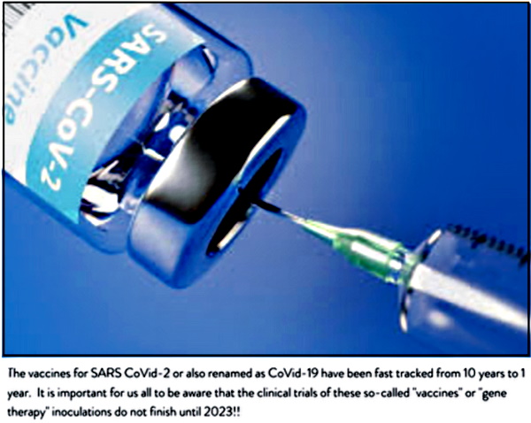
Dr. Robert Young & Science Team Reveal Graphene, Aluminum, Lipid Nanoparticle (LNP) Capsids, Poly-Ethylene Glycol (PEG) & Parasites in Pfizer, Moderna, Astrazeneca & Janssen Vaccines
See Dr. Young’s full report below the video.
Breaking: Dr. Young Reveals Graphene, Aluminium, LNP Capsids, Parasite in 4 Vaccines
by Ramona D Reports
August 27, 2021
Original video available at Ramona D Reports BitChute channel.
[As a service to protect truth from censorship and to share widely, mirrored copies of this video are available at Truth Comes to Light Odysee, BitChute and Brighteon channels. All credit, along with our sincere thanks, goes to the original source of this video. Please follow links provided to support their work.]
Scanning & Transmission Electron Microscopy Reveals Graphene Oxide in CoV-19 Vaccines
Phase Contrast Microscopy, Transmission and Scanning Electron Microscopy and Energy-Dispersive X-ray Spectroscopy Reveal the Ingredients in the CoV-19 Vaccines!
by Dr. Robert O. Young
Updated August 27, 2021

Abstract
Currently there are four major pharmaceutical companies who manufacture a SARS-CoV-2 now called SARS-CoV-19 vaccine. These manufactures and their vaccine are Pfizer–BioNTech mRNA Vaccine, the Moderna-Lonza mRNA-1273 Vaccine, the Serum Institute Oxford Astrazeneca Vaccine and the Janssen COVID -19 Vaccine, manufactured by Janssen Biotech Inc., a Janssen Pharmaceutical Company of Johnson & Johnson, a recombinant, replication-incompetent adenovirus type 26 expressing the SARS-CoV-2 spike protein. The intended purpose of these vaccines are to provide immunity from the so-called infectious novel coronavirus or SARS-CoV – 2 virus now called the SARS-CoV – 19. These four pharmaceutical companies have not provided complete FDA disclosure on their vaccine box, insert fact sheet or label for many of the major and/or minor ingredients contained within these so-called vaccines. The purpose of this research article is to identify those specific major and minor ingredients contained in the Pfizer Vaccine, the Moderna Vaccine, the Astrazeneca Vaccine and the Janssen Vaccine using various scientific anatomical, physiological and functional testing for each SARS-COV-2-19 vaccine. As a human right, governed under World Law by the Nuremberg Code of 1947, the vaccine specific ingredient information is critical, required and necessary to know so that any human from any country in the World can make an informed decision whether or not to consent to the SAR-CoV-2-19 inoculation. We have conducted the scientific testing on each vaccine and have identified several ingredients or adjuvants that have not been disclosed which are contained in these four SARS-CoV – 2 -19 vaccines. Currently, these vaccines are being administered to millions of humans around the World under an Emergency Use Authorization (EUA) issued by each country without full disclosure of all ingredients and in some cases mandated by governments or employers in violation of individual human rights under the Nuremberg Code of 1947.
Methodology and Techniques
Four “vaccines” were analyzed which are the Pfizer-BioNtech, Moderna-Lonza mRNA-1273 Vaccine, Vaxzevria by Astrazeneca, Janssen by Johnson & Johnson, using different instrumentation and protocols of preparation according to new nano particulate technological approaches. The different instrumentation includes Optical Microscopy, Bright-Field Microscopy, pHase Contrast Microscopy, Dark-Field Microscopy, UV absorbance and Fluorescence Spectroscopy, Scanning Electron Microscopy, Transmission Electron Microscopy, Energy Dispersive Spectroscopy, X-ray Diffractometer, Nuclear Magnetic Resonance instruments were used to verify the “vaccines” morphologies and contents. For the high-technology measurements and the care of the investigation, all the controls were activated and reference measurements adopted in order to obtain validated results.
Live Blood Phase Contrast and Dark-Field Microscopy
Images of the aqueous fractions of the vaccines were subsequently obtained to visually assess the possible presence of carbon particulates or graphene.
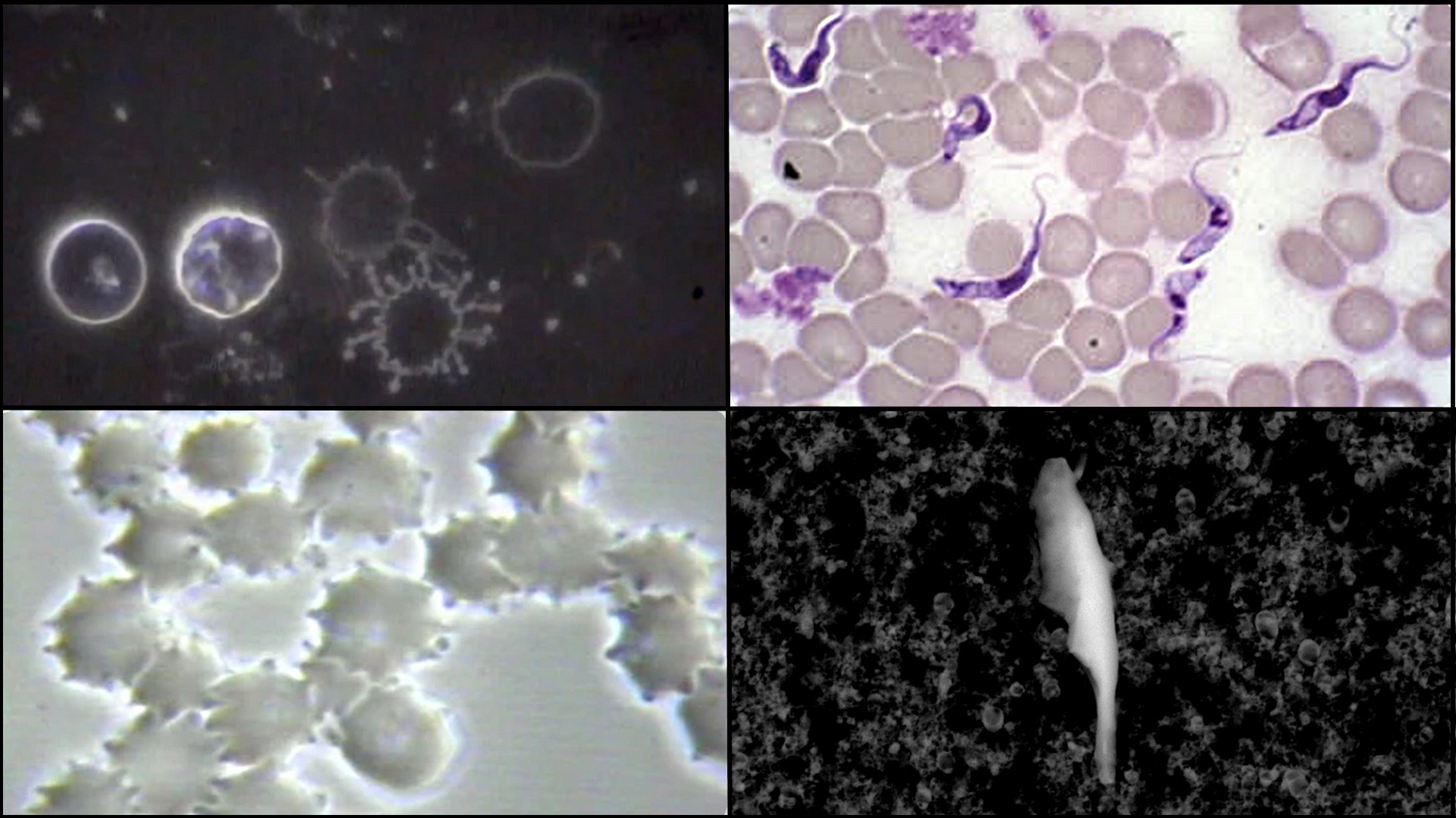
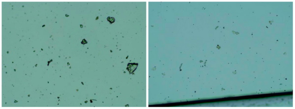
The observations under optical microscopy revealed and abundance of transparent 2D laminar objects that show great similarity with images from literature (Xu et al, 2019), and with images obtained from rGO standard (SIGMA)(Figures 1, 2 and 3).
Images of big transparent sheets of variable size and shapes were obtained, showing corrugated and flat, irregular. Smaller sheets of polygonal shapes, also similar to flakes described in literature (Xu et al, 2019) can be revealed with pHase Contrast and Dark-Field microscopy (Figure 3).
All these laminar objects were widespread in the aqueous fraction of the blood (Figure 1) or vaccine sample (Figures 2 and 3) and no component described by the registered patent can be associated with these sheets.
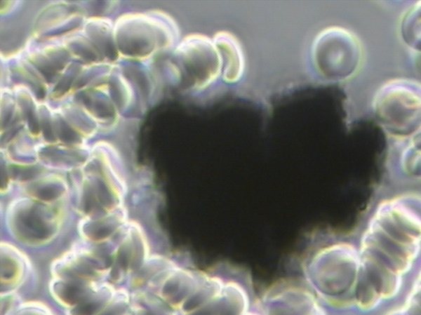
In Figure 1 You Can See What A Cluster Bomb of Reduced Graphene Oxide (rGO) Looks Like in the Live Unstained Human Blood after a CoV-19 Inoculation Causing Pathological Blood Coagulation![1][2][55][56][57]
What Are the Non-Disclosed Ingredients Contained in CoV – 19 So-Called Pfizer, Moderna, Astrazeneca and Janssen Vaccines?
To answer this question an aqueous fraction of the Pfizer, Moderna, Astrazeneca and Janssen vaccines were taken from each vile and then viewed separately under pHase Contrast Microscopy at 100x, 600x up to1500x magnification showing anatomical evidence of reduced Graphene Oxide (rGO) particulates which were compared to micrographs of rGO from Choucair et al, 2009 for identification and verification.[3]
Steps of Analysis of Vaccine Aqueous Fractions
Refrigerated samples were processed under sterile conditions, using laminar flow chamber and sterilized lab ware.
Steps for analyses were:
1. Dilution in 0.9% sterile physiological saline (0.45 ml + 1.2 ml)
2. Polarity fractionation: 1.2 ml hexane + 120 ul of RD1 sample
3. Extraction of hydrophilic aqueous pHase
4. UV absorbance and fluorescence spectroscopy scanning
5. Extraction and quantification of RNA in the sample
6. Electron and optical microscopy of aqueous pHase
The Pfizer “Vaccine” Non-disclosed Ingredients
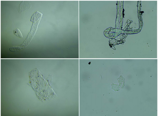
The micrographs in Figures 2 and 3 were obtained using 100X, 600X and 1500X pHase Contrast, Dark Field and Bright Field Optical Microscopy.[3]
On the left of each micrograph you will view micrographs obtained from the Pfizer vaccine aqueous fraction containing rGO.
On the right of each micrograph ou will view a match from known sources containing rGO for anatomical validation.
The observations under a pHase Contrast, Dark-Field, Bright-Field microscopy, Transmission and Scanning Electron microscopy of the vaccine product by Pfizer, including vaccine products of Moderna, Astrazeneca and Janssen revealed some entities that can be graphene strips as seen below in Figure 3.
For a definitive identification of graphene by TEM, it is necessary to complement the observation with the structural characterization by obtaining a characteristic electron diffraction standard sample (as the figure ‘b’ shown below).
The standard sample corresponding to graphite or graphene has a hexagonal symmetry, and generally has several concentric hexagons.
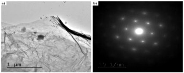
Using Transmission Electron Microscopy (TEM) we observed an intricate matrix or mesh of folded translucent flexible rGO sheets with a mixture of darker multilayer agglomerations and lighter colored of unfolded monolayers as seen in Figure 5. [3]
The darker linear areas in Figure 5 appear to be local overlap of sheets and local arrangement of individual sheets in parallel to the electron beam.[4]
After the mesh, a high density of unidentified rounded and elliptical clear shapes appears, possibly corresponding to holes generated by mechanical forcing of the rGO mesh during treatment as seen in Figure 6.[4]
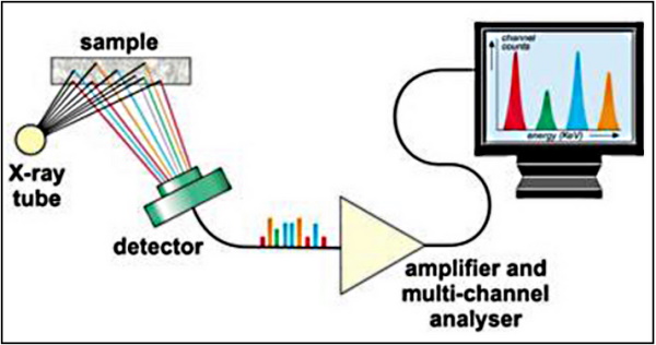
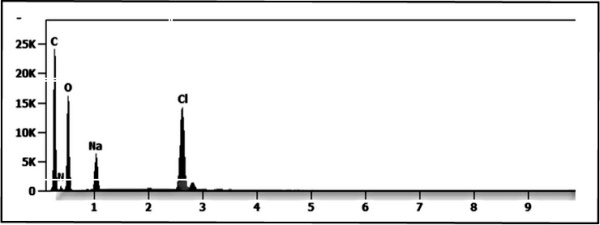
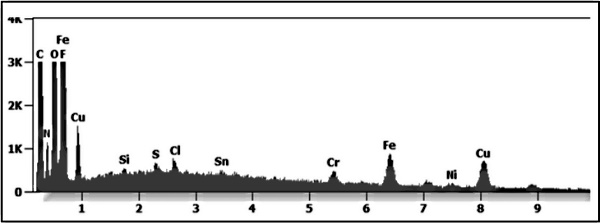
Energy-Dispersive X-ray Spectroscopy Reveals rGO in Pfizer Vaccine[5][6][7]
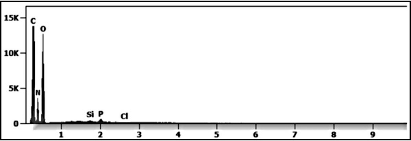
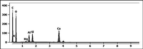
The Pfizer vaccine liquid fraction was then analyzed for chemical and elemental content using Energy-dispersive X-ray spectroscopy (EDS) as seen in Figure 6. The EDS spectrum showed the presence of Carbon, Oxygen verifying the rGO elements and Sodium and Chloride since the sample shown in Figures 2, 3, 5, and 6 were diluted in a saline solution.
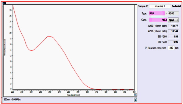
The Quantification of mRNA in the Pfizer Vaccine
The quantification of RNA in the Pfizer sample was carried out with conventional protocols (Fisher).
According to NanoDropTM 2000 spectrophotometer calibration check specific software (Thermofisher), the UV absorption spectrum of total aqueous fraction was correlated to 747 ng/ul of unknown absorbing substances.
However, after RNA extraction with commercial kit (Thermofisher), quantification with RNA specific Qbit fluorescence probe (Thermofisher) showed that only 6t ug/ul could be related to the presence of RNA. The spectrum was compatible with the peak of rGO at 270nm.
According to microscopic images presented here, most of this absorbance might be due to graphene-like sheets, abundant in the fluids suspension in the sample.
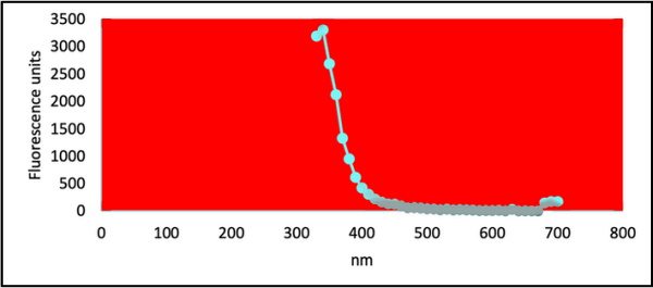
The conclusions are further supported by high fluorescence from the sample with maximum at 340 nm, in accordance with peak values for rGO. It must be reminded that RNA does not show spontaneous fluorescence under UV exposure.
Ultra Violet Fluorescence Testing of the Pfizer Aqueous Fraction for Reduced Graphene Oxide (rGO)[5]
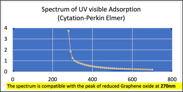
Ultra Violet absorption and fluorescence spectra were obtained with Cytation 5 Cell Imaging Multi-Mode Reader Spectrophotometer (BioteK). UV absorbance spectrum confirmed a maximum peak at 270nm, compatible with presence of rGO particulate.
UV fluorescence maximum at 340 nm also suggests presence of significant amounts of rGO in the sample (Bano et al, 2019).
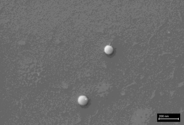
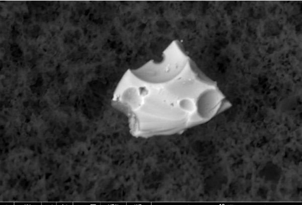
Figures 11 and 12 below shows a micrograph of different micro and nano particulates which have been identified in the Pfizer, Moderna, Astrazeneca and Janssen, so-called “vaccines” and analyzed under an Environmental Scanning Electron Microscope (SEM) coupled with an x-ray microprobe of an Energy Dispersive System (EDS) that reveals the particle size, composition distribution and chemical nature of the observed micro and nano particulates under observation.[5][6][7]

Figures 13 and 14 below shows a micrograph of different micro and nano particulates which have been identified in the Pfizer, Moderna, Astrazeneca and Janssen, so-called “vaccines” and analyzed under an Environmental Scanning Electron Microscope (SEM) coupled with an x-ray microprobe of an Energy Dispersive System (EDS) that reveals the particle size, composition distribution and chemical nature of the observed micro and nano particulates under observation.
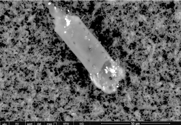
Are There Parasites in the Pfizer “Vaccines”
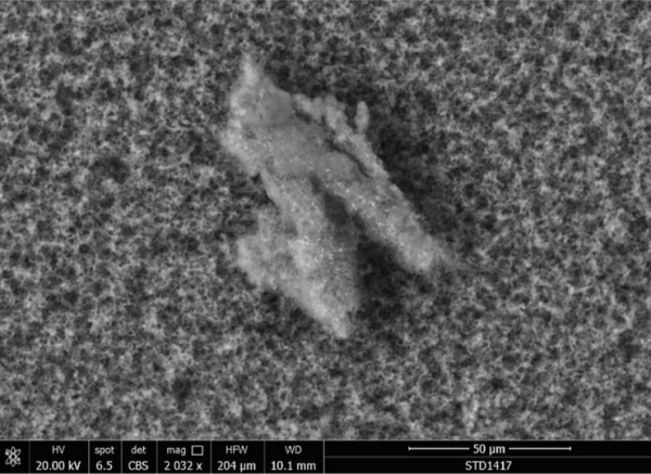
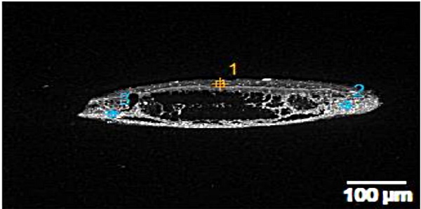
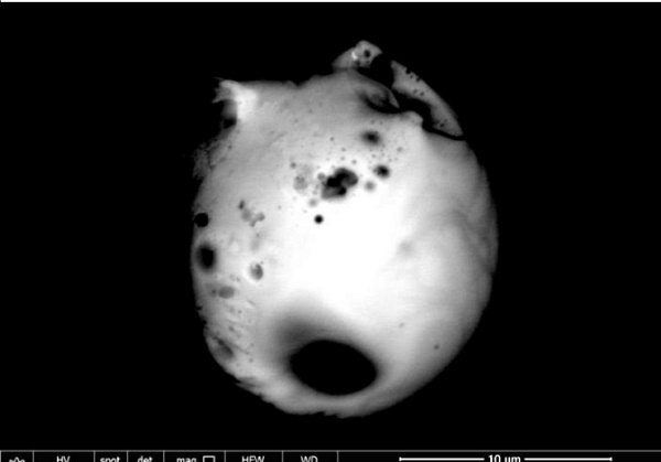
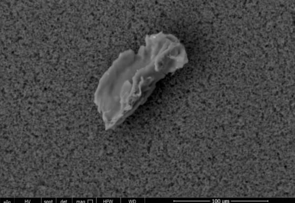
A 50 micron elongated body, as seen in Figure 13 is a sharp mysterious presence in the Pfizer vaccine. It appears and is identified anatomically as a Trypanosoma cruzi parasite of which several variants are lethal and is one of many causes of acquired immune deficiency syndrome or AIDS. [Atlas of Human Parasitology, 4th Edition, Lawrence Ash and Thomas Orithel, pages 174 to 178][8]
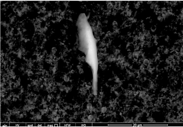
Figures 15 and 16 below show a micrograph of different micro and nano particulates which have been identified and analyzed under an Environmental Scanning Electron Microscope (SEM) coupled with an x-ray microprobe of an Energy Dispersive System (EDS) that reveals the chemical nature of the observed micro and nano particulates and their morphology.
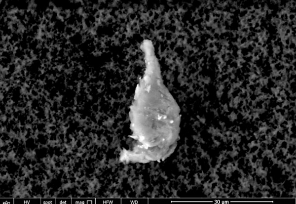
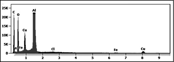
The white 2-micron-long particulate is composed of bismuth, carbon, oxygen, aluminum, sodium, copper and nitrogen.
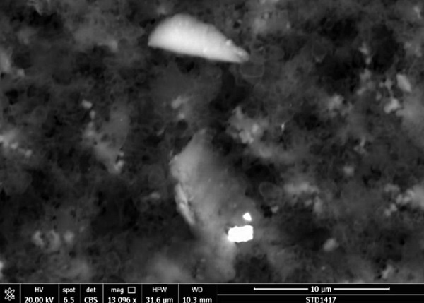
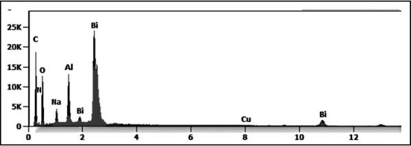
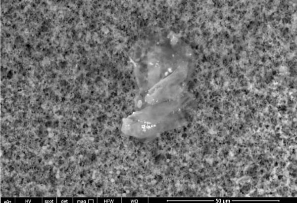
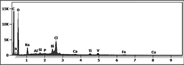
Figures 17 and 18 show the identification of organic carbon, oxygen and nitrogen particulates with an aggregate of embedded nanoparticles including bismuth, titanium, vanadium, iron, copper, silicon and aluminum which were all found in the so-called Pfizer “vaccine.”
The Astrazeneca “Vaccine” Non-disclosed Ingredients
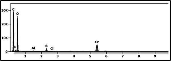
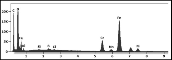
Figures 19 and 20 show an engineered aggregate of iron, chromium and nickel also known as stainless steel of micro and nano particles embedded and identified in the Astrazeneca “vaccine” viewed under Transmission Electron Microscopy (TEM) and quantified with an x-ray microprobe of an Energy Dispersive System that reveals the chemical nature of the observed micro and nano particulates and their morphology.
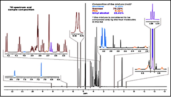
Using the XRF (X-ray fluorescence) instrument was used to evaluate the adjuvants in the Astrazeneca “vaccine”, which identified the following molecules of histidine, sucrose, Poly-ethylene glycol (PEG) and ethylene alcohol, also contained in the Pfizer and Moderna “vaccines”. The results of this test can be seen in Figure 20.[9]
The injection of PEG and Ethylene alcohol are both known as carcinogenic and genotoxic.[9] PEG was the only adjuvant declared on the data sheet listing the ingredients of the Astrazeneca “vaccine” but contained in the Pfizer and Moderna “vaccines”.
The Janssen “Vaccine” Non-Disclosed Ingredients
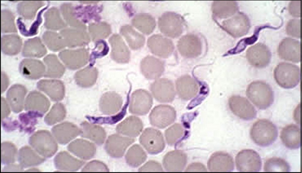
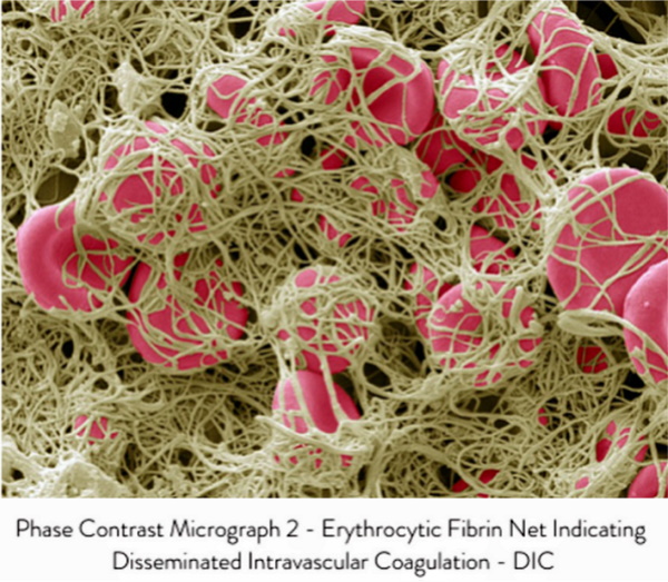
Figures 22 and 23 shows an organic-inorganic aggregate identified in the Janssen “vaccine”. The particles are composed of stainless steel and are glued together with a “Carbon-based glue” of reduced graphene oxide.[10] This aggregate is highly magnetic and can trigger pathological blood coagulation and “The Corona Effect” or “The Spike Protein Effect” creation from the degeneration of the cell membrane due to interactions with other dipoles.[10] You can view these biological reactions or cellular transformations in the live blood under pHase Contrast and Dark Field Microscopy in Figures 24, 25 and 26.[1][11]
The Corona Effect and Spike Protein Effect
The Endogenously Created “Corona Effect” and “Spike Protein” ARE Caused by Chemical and Radiation Poisoning from Reduced Graphene Oxide and Microwave Radiation![11]
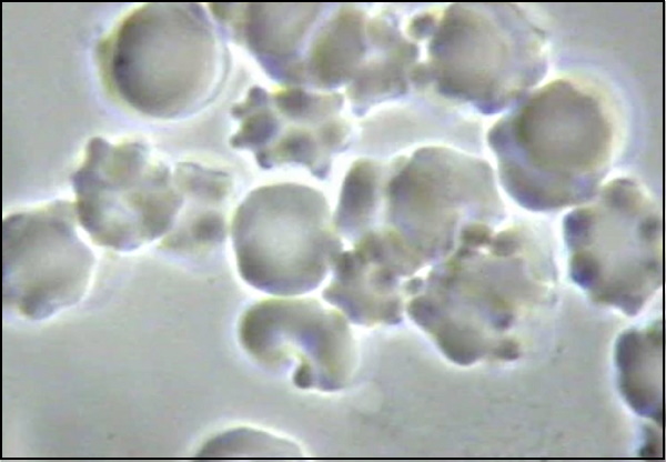
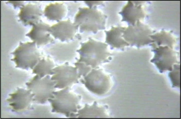
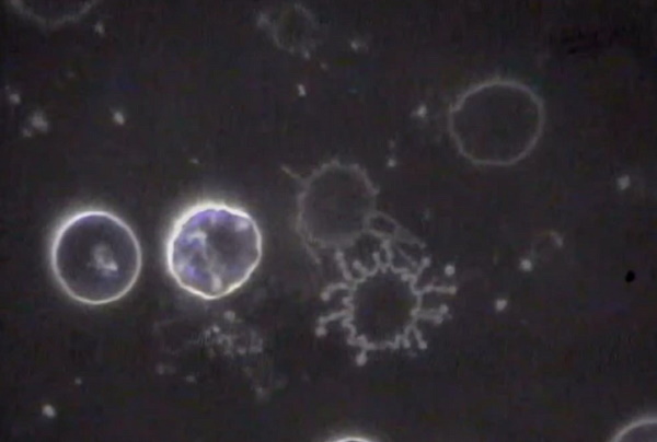
Figures 24 and 25 above show ‘The CORONA EFFECT’ on the red blood cells with Figure 26 showing ‘The SPIKED PROTEIN EFFECT’ both caused by decompensated acidosis of the interstitial and then vascular fluids from an acidic lifestyle and specifically, exposure to toxic pulsating electro-magnetic fields at 2.4gHz or higher, chemical poisoning from the food and water ingested, toxic acidic air pollution, chem-trails and to top-it-all-off a nana particulate chemical laden CoV – 19 inoculation! Please check your feelings and false beliefs at the door before YOU prematurely cause YOURSELF harm![11]
The Moderna “Vaccine” Non-Disclosed Ingredients
Figure 26 and 27 identified a mixed entity of organic and inorganic matter contained in the Moderna “vaccine.”
Transmission Electron Microscopy (TMS) and quantified with an x-ray microprobe of an Energy Dispersive System (EDS) revealed the chemical nature of the observed micro and nano particulates.
The so-called Moderna “vaccine’ is a carbon-based Reduced Graphene Oxide substrate where some nanoparticles are embedded. The nanoparticles are composed of carbon, nitrogen, oxygen, aluminum, copper, iron and chlorine.[12]
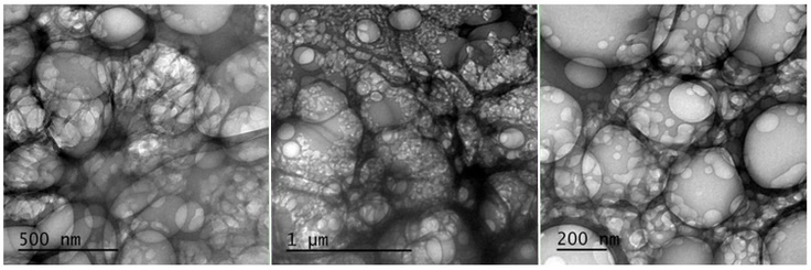
Figures 27 and 28 shows an analysis which was also performed under Transmission Electron Microscopy (TEM) and quantified with an x-ray microprobe of an Energy Dispersive System (EDS) and revealed the chemical nature of the observed micro and nano particulates. Many foreign bodies were identified with a spherical morphology with some bubble-shaped cavities.
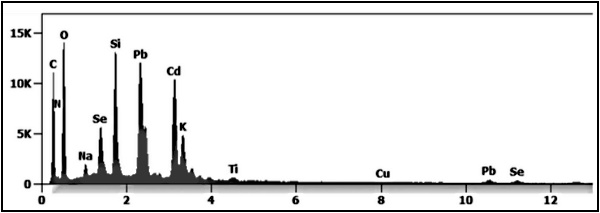
Figure 29 shows they are composed of carbon, nitrogen, oxygen, silicon, lead, cadmium, and selenium. This highly toxic nano particulate composition are quantum dots of cadmium selenide which are cytotoxic and genotoxic.[13][14]
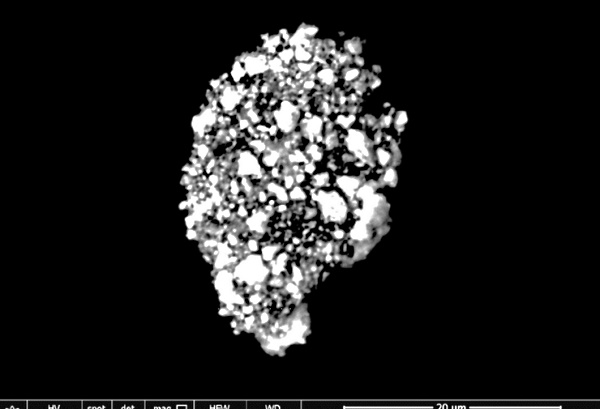
Figures 30 and 31 further analysis of the so-called Moderna “vaccine” showed a 100-micron symplast of reduced graphene oxide nano particulate composite. The rGO is composed of carbon and oxygen with contamination of nano particulates of nitrogen, silicon, phosphorus and chlorine Chlorine.[15]
Figures 32 and 33 show carbon-based reduced graphene oxdie entities in the Moderna “vaccine” mixed with aggregates filled with Aluminium silicate nanoparticulates.[16]
Discussion
The SARS-CoVid-2-19 pandemic induced the pharmaceutical industries to develop new drugs that they called vaccines.
The mechanism of action of these new drugs as declared by the pharmaceutical industry coupled with what is reported in the vaccine products’ data sheet is NOT clear for current medical savants to understand that those new drugs produced by Pfizer–BioNTech mRNA Vaccine, the Moderna-Lonza mRNA-1273 Vaccine, the Serum Institute Oxford Astrazeneca Vaccine and the Janssen COVID -19 Vaccine, manufactured by Janssen Biotech Inc., a Janssen Pharmaceutical Company of Johnson & Johnson are NOT vaccines but nanotechnological drugs working as a genetic therapy.
The name “vaccine” is likely to be an escamotage (trickery) used for bureaucratic and technocratic reasons in order to receive an urgent approval, ignoring all the normal rules necessary for new drugs, especially for those involving novel nanotechnological mechanisms which have never been developed nor experienced by humans any where, at any time in the history of World.
All these so-called “vaccines” are patented and therefore their actual content is kept secret even to the buyers, who, of course, are using taxpayers’ money. So, consumers (taxpayers) have no information about what they are receiving in their bodies by inoculation. Humanity is kept in the dark as far as the nano particulate technological processes involved are concerning, on the negative effects on the cells of the body, but mostly on the possible magneticotoxic, cytotoxic and genotoxic nano-bio-interaction effect on the blood and body cells.
This current research study via direct analysis on the aforementioned so-called “vaccines” by means of nano particulate technological instrumentation reveals disturbing and life-altering information concerning the truth about the actual toxic acidic contents of the so-called vaccines.
The Pfizer, Moderna, Astrazeneca and Janssen drugs are NOT “vaccines” but complexed Graphene Oxide nano particulate aggregates of varying nano elements attached to genetically modified nucleic acids of mRNA from animal or vero cells and aborted human fetal cells as viewed and described above. Once again the ingredients in these so-called vaccines are highly magneticotoxic, cytotoxic and genotoxic to plant, insect, bird, animal and human cell membranes and their genetics which already has lead to serious injuries (estimated at over 500 million) and/or eventual death (estimated at over 35 million).[17][18] through [54]
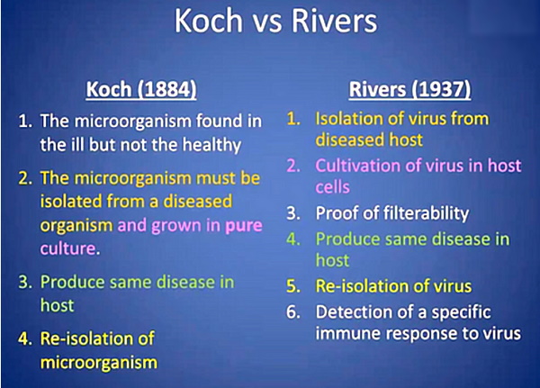
The so-called “experts” or “medical savants” are telling YOU that CoV -2 – 19 vaccines are the ONLY way to stop the spread of CoV-19… even when there is NO EVIDENCE of its existence and NO EVIDENCE of it spreading as determined by the scientific method of Koch or Rivers postulates![53]
That they’re safe — despite the documented evidence is to the contrary…[53]
That they’re effective — even though millions of “double-jabbed” people are getting sick, theoretically exposing themselves to a NON-EXISTENT VIRUS called CoV – 19, and dying…[54] NOT from some phantom viral infection but from the FEAR or false evidence appearing real and the toxic acid contents of reduced graphene oxide delivered via the genetically modified mRNA to specific targets of the human body leading to pathological blood coagulation, oxygen deprivation, hypercapnia, hypoxia and then death by suffocation.[55][56][57]
That YOU MUST get at LEAST two shots PLUS “boosters” to live “normal lives”…
And soon, they’ll be telling YOU that YOU have no choice but to comply with ALL their MANdates even when the CDC and other Governments, Universities and Medical Institutes have admitted in writing that they have NO “GOLD STANDARD” isolation of the CoV – 2 now called CoV – 19 virus![54]
There is NO CORONA VIRUS and NEVER HAS BEEN![55]
Remember …
DON’T LET ANYONE TAKE AWAY YOUR HEALTH FREEDOM!
It is YOUR Body, YOUR Life and YOUR Choice!
Knowledge is power. And it’s the key to understanding why the experimental CoV -19 vaccines are so dangerous — despite the corporate media’s official narrative that suppresses and censors anyone who dares to speak out.
You are in control of your own health. Don’t fall victim to global governments and bureaucrats that are pushing everyone to get vaccinated. Billionaire “philanthropist” Bill Gates and billionaire Big Tech activists think they know what’s best for you and your family.
You must be free to decide what’s right for you. Do NOT let governments and employers force you into getting “vaxxed” “for your own good”.
And never let the cancel culture make you too afraid to stand up for your rights!
In the words of the great French doctor and scientist, Antione BeChamp, “there is nothing so false that does NOT contain a element of truth and so it is with the germ theory.” In this case the viral, vaccine and immunity theory![58]
Connect with Dr. Robert O. Young
See related:
Dr. Andrew Kaufman w/ SGT Report: Something Wicked This Way Comes — On the Hidden Agenda Behind, and Hidden Ingredients Within, the Toxic Covid “Vaccines”


Truth Comes to Light highlights writers and video creators who ask the difficult questions while sharing their unique insights and visions.
Everything posted on this site is done in the spirit of conversation. Please do your own research and trust yourself when reading and giving consideration to anything that appears here or anywhere else.